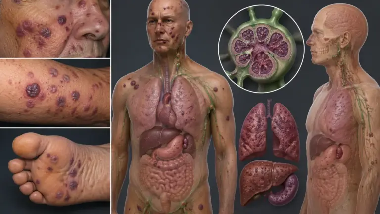
📌 What You Need to Know in a Nutshell
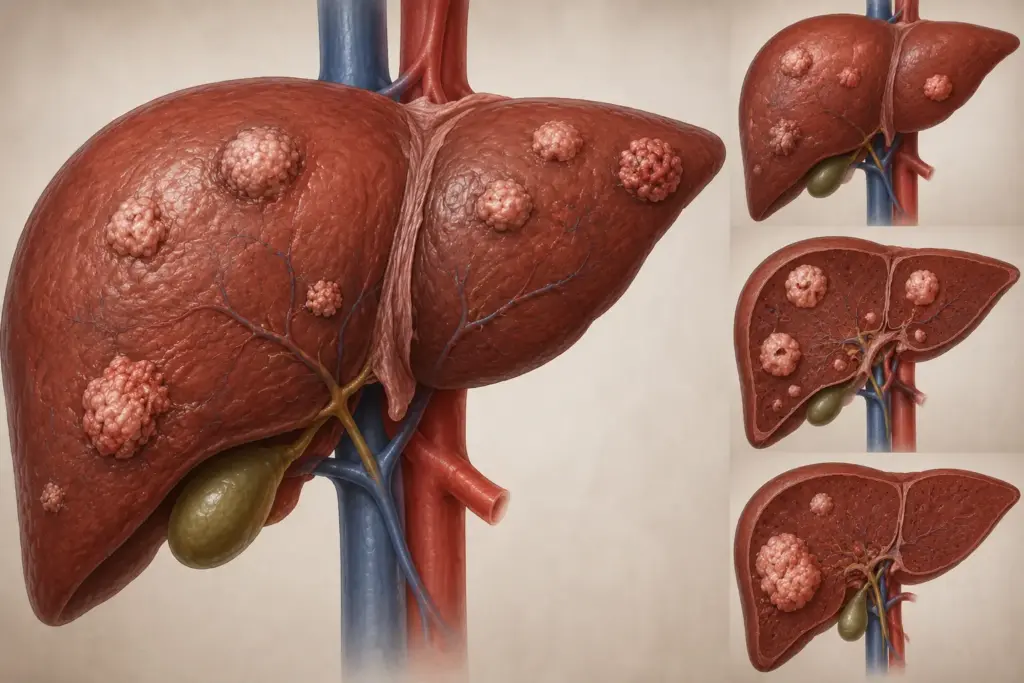
- Liver cancer, is classified into two main groups: primary tumors, known as hepatocellular carcinoma (HCC), and metastatic tumors; primary HCC is the most common primary liver malignancy in adults.
- The 5-year survival rate for localized disease is approximately 38%; in small tumors that can be completely resected surgically, this rate can reach 90%. In the metastatic stage, the 5-year survival rate is approximately %4.
- In early stages (BCLC 0-A), surgical resection or ablation is the primary systemic treatment, while in advanced stages, immune checkpoint inhibitors (atezolizumab + bevacizumab) are the primary systemic treatment.
- The hospital stay following liver cancer surgery (hepatectomy) is 5–10 days; full recovery occurs within 6–8 weeks.
Liver cancer is a heterogeneous group of diseases that includes primary liver malignancies—primarily hepatocellular carcinoma (HCC) primarily, is a heterogeneous group of diseases encompassing primary liver malignancies and metastatic liver tumors. In 2020, 905,677 new cases of HCC were reported worldwide, and this disease was the third leading cause of cancer deaths. A distinctive feature of the diseaseis that it is asymptomatic in the early stages and most often develops in livers with cirrhosis; this can both delay diagnosis and limit treatment options.
What Is Liver Cancer?
Liver cancer is the general term for malignant neoplasms originating in liver tissue. Among primary liver cancers, hepatocellular carcinoma (HCC) is the most common histological type, accounting for 75–85% of cases. Intrahepatic cholangiocellular carcinoma and combined hepatocellular-cholangiocellular carcinoma are less common. Approximately 80–90% of HCCs develop in livers with cirrhosis; therefore, patients with chronic liver disease are at high risk.
According to World Health Organization data, HCC was recorded as the third leading cause of cancer deaths worldwide in 2020, with 905,677 new cases and 830,180 deaths. The disease is 2 to 4 times more common in men than in women. The strongest risk factors include chronic hepatitis B (HBV) and hepatitis C (HCV) infections, alcoholic cirrhosis, nonalcoholic fatty liver disease (NAFLD), and exposure to aflatoxins.
According to Assoc. Prof. Dr. Nilay Şengül, the vast majority of patients diagnosed with liver cancer have an underlying chronic liver disease; this necessitates that not only the tumor but also liver function be taken into account when planning treatment. In recent years, systemic treatment outcomes have improved significantly with immune checkpoint inhibitors and targeted therapies.
Symptoms of Liver Cancer
Symptoms of liver cancer vary depending on the stage of the disease and underlying liver function. The disease, which is generally asymptomatic in the early stages, leads to specific and nonspecific symptoms as it progresses. The table below :
| Stage | Typical Symptoms | Liver Function |
|---|---|---|
| Early (BCLC 0-A) | Usually asymptomatic; detected incidentally by ultrasound or AFP screening | Child-Pugh A (compensated) |
| Intermediate (BCLC B) | Right upper quadrant pain, fatigue, loss of appetite, weight loss, abdominal distension (mild ascites) | Child-Pugh A-B |
| Advanced (BCLC C) | Abdominal distension (ascites), jaundice, itching, nausea and vomiting, enlarged liver (hepatomegaly) | Child-Pugh B-C |
| Terminal (BCLC D) | Confusion (hepatic encephalopathy), bleeding (variceal bleeding), kidney failure, severe jaundice | Child-Pugh C (decompensated) |
Stage 1 Liver Cancer Symptoms
Symptoms of stage 1 liver cancer are generally absent. The disease is usually detected incidentally during routine ultrasound and alpha-fetoprotein (AFP) measurements. Rarely, patients may report mild discomfort or a sensation of fullness in the right upper abdomen. At this stage, diagnosis is the most critical factor in significantly improving the patient’s long-term survival.
Stage 4 Liver Cancer Symptoms
Stage 4 liver cancer symptoms indicate the terminal stage of the disease (BCLC D) and include the following:
- Jaundice in the final stage of liver cancer: When a tumor or tumor thrombus in the liver blocks the bile ducts, jaundice (icterus) develops in the skin and the whites of the eyes. Bilirubin levels rise significantly.
- Liver cancer-related fluid accumulation in the abdomen (ascites): Fluid accumulates in the abdominal cavity as a result of portal hypertension and liver failure. This causes significant abdominal swelling, shortness of breath, and a feeling of early satiety.
- Confusion and drowsiness (hepatic encephalopathy): When the liver is unable to eliminate ammonia and other toxic substances, it can lead to impaired consciousness, confusion, and coma.
- Tendency to bleed: Due to thrombocytopenia and a deficiency in clotting factors, nosebleeds, bleeding gums, or gastrointestinal bleeding may occur.
Symptoms of end-stage liver cancer also include severe cramping pain, edema, kidney failure, and cachexia (severe weight loss). Symptoms of death in stage 4 liver cancer are generally associated with hepatic encephalopathy, variceal bleeding, or hepatorenal syndrome.
Assoc. Prof. Dr. Şengül emphasizes that a significant proportion of patients diagnosed with liver cancer in clinical practice present at an advanced stage because the disease does not present symptoms in the early stages. For this reason, she notes that six-monthly ultrasound and AFP monitoring are of vital importance in patients with chronic liver disease.
What Causes Liver Cancer?
The causes of liver cancer are mostly associated with chronic liver disease and cirrhosis. The list below summarizes the primary risk factors proven in the scientific literature:
- Chronic Hepatitis B (HBV): Approximately 50% of HCC cases worldwide are associated with HBV. The annual risk of HCC in chronic carriers is 0.3–0.6%; in those who develop cirrhosis, this risk rises to 2.5–3.6%.
- Chronic Hepatitis C (HCV): The annual risk of HCC in HCV patients who develop cirrhosis is 1–4%. Eradication of HCV through antiviral therapy significantly reduces the risk of HCC.
- Alcoholic Cirrhosis: Heavy alcohol consumption increases the risk of cirrhosis and HCC. The combination of alcohol and HBV/HCV synergistically increases this risk.
- Nonalcoholic Fatty Liver Disease (NAFLD/NASH): Along with the obesity and type 2 diabetes pandemics, it has become one of the leading emerging risk factors for HCC.
- Aflatoxin Exposure: Aflatoxin B1, produced by Aspergillus fungi, can increase the risk of HCC by up to 30-fold in HBV carriers.
- Metabolic Syndrome: The combination of obesity, diabetes, dyslipidemia, and hypertension increases the risk of NAFLD and, consequently, HCC.
Other factors contributing to liver cancer include hemochromatosis, alpha-1 antitrypsin deficiency, Wilson’s disease, and primary sclerosing cholangitis. Regular HCC screening is recommended for all of these conditions.
Stages of Liver Cancer
Liver cancer stages are most commonly classified using the Barcelona Clinic Liver Cancer (BCLC) system. This system evaluates tumor burden, liver function (Child-Pugh score), performance status (ECOG), and symptoms. The AASLD and EASL guidelines recommend the BCLC system.
| BCLC Stage | Definition | Treatment | Median Survival |
|---|---|---|---|
| 0 (Very Early) | Single nodule <2 cm, Child-Pugh A | Ablation, resection, transplant | >5 years |
| A (Early) | 1–3 nodules ≤3 cm or single nodule ≤5 cm, Child-Pugh A–B | Resection, transplant, ablation | >5 years |
| B (Intermediate) | Multiple nodules, Child-Pugh A-B, ECOG 0 | TACE (transarterial chemoembolization) | >2.5 years |
| C (Advanced) | Portal vein invasion, extrahepatic spread, ECOG 1–2 | Systemic therapy | >2 years |
| D (Terminal) | Child-Pugh C or ECOG >2 | Palliative care | ~3 months |
AFP levels are also a prognostic factor in determining liver cancer stages. According to a study published in PMC, the median survival for patients with BCLC Stage A and AFP ≥200 ng/mL is 28 months, it was 39 months for those with AFP <200 ng/mL. In BCLC Stage B, this difference is more pronounced: 19 months for AFP ≥200 ng/mL and 22 months for <200 ng/mL.
The survival time for stage 3 liver cancer is, on average, over 2 years for BCLC stage C; however, this duration may vary depending on the patient’s liver function, ECOG performance status, and response to treatment. The average survival time for stage 4 liver cancer (BCLC D) is approximately 3 months.
Liver Cancer Treatment
Liver cancer treatment is personalized based on the BCLC stage, liver function, tumor characteristics, and the patient’s overall condition. The treatment approach ranges from curative to palliative.
Liver Cancer Treatment Methods
1. Surgical Resection (Hepatectomy): It is performed for curative purposes in patients with BCLC stages 0 and A who have sufficient future liver reserve (FLR) and compensated cirrhosis. According to the AASLD guidelines, the absence of portal hypertension and sufficient FLR are required in patients with cirrhosis. Minimally invasive (laparoscopic/robotic) approaches can reduce perioperative morbidity.
2. Liver Transplantation: It is the gold standard treatment for patients who meet the Milan criteria (single tumor ≤5 cm or 2–3 tumors ≤3 cm). The 5-year survival rate after transplantation is 75% or higher. The AASLD recommends bridging therapy prior to transplantation (TACE, TARE, ablation).
3. Local Ablative Therapies: Radiofrequency ablation (RFA) and microwave ablation (MWA) are used as alternatives to surgery for early-stage tumors ≤3 cm. The AASLD recommends thermal ablation as an alternative to surgery .
4. Transarterial Chemoembolization (TACE): This is the standard treatment for BCLC Stage B (intermediate stage). The tumor’s blood supply is cut off by administering a tumor-specific chemotherapy drug and an embolizing agent. Radioembolization (TARE) can be used as an alternative.
5. Systemic Therapy: This is the preferred treatment for BCLC Stage C. The atezolizumab + bevacizumab combination was approved as first-line therapy based on the results of the IMbrave150 trial. In the HIMALAYA trial, the durvalumab + tremelimumab combination demonstrated superiority over sorafenib (median OS 16.4 vs. 13.7 months). Lenvatinib is an alternative for patients who are not eligible for immunotherapy.
6. Second- and Third-Line Treatments: Cabozantinib, regorafenib, and ramucirumab (in patients with AFP ≥400 ng/mL) and the nivolumab + ipilimumab combination can be used in advanced-stage disease.
According to Assoc. Prof. Dr. Nilay Şengül, a multidisciplinary approach is of vital importance in the treatment of liver cancer; joint decision-making by specialists in surgery, medical oncology, radiation oncology, and gastroenterology specialists working together to make decisions optimizes both treatment selection and patient outcomes. In particular, with the introduction of immune checkpoint inhibitors into systemic therapy, survival in advanced HCC has been significantly prolonged.
| BCLC Stage | First-Line Treatment | Alternative/Second-Line |
|---|---|---|
| 0-A | Surgical resection or transplantation | RFA/MWA (≤3 cm, if not a candidate for surgery) |
| B | TACE | TARE, SBRT |
| C | Atezolizumab + bevacizumab or durvalumab + tremelimumab | Lenvatinib, sorafenib (if immunotherapy is contraindicated) |
| D | Palliative care, symptom management | Lenvatinib or sorafenib may be tried in some cases |
Liver Cancer Surgery
Liver cancer surgery (hepatectomy) is the primary surgical procedure performed for curative intent in BCLC stages 0–A. During the procedure, liver segments containing the tumor are removed; a right hemihepatectomy (segments 5–8), a left hemihepatectomy (segments 1–4), or a segmentectomy may be performed. A calculation of the functional liver reserve (FLR) must be performed during the preoperative period.
Surgical approaches are grouped into three main categories:
- Open Hepatectomy: This is the traditional approach; it may be preferred for large tumors and complex resections. The average duration of the procedure is 3–6 hours.
- Laparoscopic Hepatectomy: This is a minimally invasive approach; it results in less pain, a shorter hospital stay, and faster recovery. The AASLD recommends the minimally invasive approach for selected patients.
- Robot-Assisted Hepatectomy: It offers advantages similar to laparoscopy; it allows for precise dissection, particularly in confined spaces.
The answer to the question “How long does liver cancer surgery take?” varies depending on the extent of the resection. Minor resections (2 segments or fewer) take 2–3 hours, while major resections (3 or more segments) 3–6 hours. According to a study published in PMC, the duration of the operation was found to be an independent predictor of morbidity.
Risks of Liver Cancer Surgery
The risks of liver cancer surgery vary depending on liver function, the extent of resection, and comorbidities. According to a comprehensive study published in PMC, the morbidity rate was reported as 39%, and the 90-day mortality rate as 5.2%. The most common complications are as follows:
- Post-hepatectomy liver failure (PHLF): This is the most serious complication; it occurs in approximately 10% of resections. It is characterized by an increase in INR and hyperbilirubinemia. High preoperative total bilirubin levels and major resection increase the risk.
- Bile leak: It can occur in 4–17% of resections; most cases are managed conservatively or with drainage.
- Bleeding: Intraoperative or postoperative bleeding; more common in major resections.
- Pleural effusion and pneumonia: Subphrenic infections and respiratory complications.
- Surgical site infection: Surgical site infections.
The answer to the question “How long do patients who undergo liver cancer surgery live?” depends on the stage of the disease and its pathological characteristics. In localized HCC, the 5-year survival rate after surgical resection is between 50% and 70%; in small tumors that can be completely removed, this rate can reach 90%. However, the risk of recurrence is high in livers with cirrhosis; therefore, adjuvant therapy and close follow-up may be necessary.
Assoc. Prof. Dr. Şengül emphasizes that the most critical period following liver resection in clinical practice is the first 5 days, because-hepatectomy-related liver failure typically occurs during this period. She notes that careful evaluation of preoperative liver function and calculation of adequate future liver reserve significantly reduce mortality and morbidity.
Clinical Assessment by Assoc. Prof. Dr. Nilay Şengül
In my clinical practice, I frequently observe that approximately 60–70% of patients diagnosed with liver cancerpresent at stages BCLC B, C, or D; this is related to the disease being asymptomatic in its early stages and the inadequacy of screening programs.
According to Assoc. Prof. Dr. Nilay Şengül, the most critical factor for success in liver cancer treatment is early diagnosis; 6-monthly ultrasound and AFP monitoring in chronic hepatitis B carriers and patients with cirrhosis significantly increases the chances ofsignificantly increases the chances of detecting HCC at an early stage and achieving curative treatment. In recent years, with the introduction of immune checkpoint inhibitors into systemic therapy, median survival in advanced-stage HCC has increased from 13–16 months to over 20 months; this development has further underscores the importance of patient-specific treatment strategies.
From Diagnosis to Recovery: How Does the Process Proceed?
- Initial Examination and Evaluation: Detailed information is obtained from the patient regarding a history of liver disease (HBV, HCV, alcohol, NAFLD), family history, and symptoms (pain, weight loss, jaundice, ascites). During the physical examination, liver enlargement, ascites, splenomegaly, and jaundice are assessed.
- Laboratory Tests: Complete blood count, liver function tests (AST, ALT, ALP, GGT, total bilirubin, albumin), INR, creatinine, AFP, and AFP-L3% are measured. An AFP level >200 ng/mL supports a diagnosis of HCC; an AFP-L3% ≥ 10% indicates a high risk of HCC.
- Imaging: Tumor characterization is performed using contrast-enhanced multi-phase liver CT or contrast-enhanced MRI. HCC is defined by a hypervascular lesion in the arterial phase and wash-out in the venous/late phase. Chest CT is used for metastasis screening. The AASLD does not recommend the routine use of PET or bone scintigraphy.
- Pathological Confirmation: In cases where a diagnosis cannot be made with imaging, percutaneous biopsy or biopsy guided by EUS (endoscopic ultrasound) may be performed. The risk of implantation should be considered prior to biopsy.
- Staging and Treatment Planning: BCLC staging, Child-Pugh score, and ECOG performance status are determined. Specialists in surgery, medical oncology, radiation oncology, gastroenterology, and radiology meet in a multidisciplinary tumor board (MDT).
- Treatment Implementation: If the patient is a candidate for surgery, resection or transplantation is planned. In the intermediate stage, TACE is performed. In the advanced stage, systemic therapy (atezolizumab + bevacizumab or durvalumab + tremelimumab) is initiated.
- Hospital Stay and Discharge: The hospital stay following hepatectomy is 5–10 days. Discharge criteria include tolerance of oral nutrition, pain control, adequate urine output, absence of complications, and stable liver function.
- Follow-up and Recovery: The first follow-up visit is scheduled 1–2 weeks later. AFP and imaging follow-up continue every 3–6 months. Because the risk of recurrence is high, close monitoring is recommended for the first 2 years. Return to work is generally possible within 6–8 weeks.
Frequently Asked Questions
How Is Liver Cancer Diagnosed?
It is diagnosed using contrast-enhanced multi-phase liver CT or MRI. HCC is characterized by hypervascularity in the arterial phase and wash-out in the venous/late phase-out in the venous/late phase. An AFP level >200 ng/mL supports the diagnosis. In cases where a diagnosis cannot be made with imaging, a biopsy may be performed.
Is an Enlarged Liver Cancer?
No, an enlarged liver (hepatomegaly) is not always cancer. Cirrhosis, fatty liver, hepatitis, congestive heart failure, and benign tumors can also cause hepatomegaly. A definitive diagnosis is made through imaging and, if necessary, a biopsy.
How Is Liver Cancer Diagnosed?
It is generally not detectable in the early stages. Early diagnosis is possible in patients with chronic liver disease through ultrasonography and AFP screening every 6 months. In advanced stages, it presents with symptoms such as abdominal pain, weight loss, jaundice, ascites, and an enlarged liver.
What Does Liver Cancer Pain Feel Like?
It presents as constant or intermittent pain in the upper right abdomen, below the ribs. Spread to the tumor capsule or invasion of the abdominal wall can intensify the pain. Rarely, it can lead to shoulder pain (phrenic nerve involvement).
What Helps with Liver Cancer?
Treatment selection is determined by the stage of the disease. In early stages, surgery or ablation may be curative. In advanced stages, immune checkpoint inhibitors and targeted therapies help control the disease. Alcohol should be completely avoided, and a balanced diet and regular exercise are recommended.
What Should You Eat If You Have Liver Cancer?
A low-sodium, high-protein, and balanced diet is recommended. If cirrhosis is present, sodium restriction (<2 g per day) helps prevent acid formation. Caffeine consumption may reduce the risk of HCC in chronic liver disease. According to the AASLD guidelines, coffee consumption may be recommended. Raw seafood and foods containing aflatoxins should be avoided.
What Is the AFP Level in Liver Cancer?
In healthy adults, the normal AFP level ranges from 0 to 8 ng/mL. In chronic liver disease, an AFP level >200 ng/mL supports a diagnosis of HCC. Very high levels (500–1,000 ng/mL and above) are generally considered a sign of cancer. An AFP-L3% ≥ 10% indicates a high risk of HCC.
Can Liver Cancer Be Detected Through a Blood Test?
Yes, HCC can be detected with an AFP blood test. However, elevated AFP levels do not always indicate HCC; they can also be elevated in cases of acute hepatitis, cirrhosis, and pregnancy. AFP alone is not diagnostic; it must be evaluated in conjunction with imaging.
Can Liver Cancer Be Detected in the Blood?
It may be partially detectable. Serum markers such as AFP, PIVKA-II, and AFP-L3 support the diagnosis. However, a definitive diagnosis is made through imaging (CT/MRI). Elevated levels of these biomarkers in the blood are used to monitor the disease and for the early detection of recurrence.
Is There a Treatment for Liver Cancer?
Yes, curative treatment is possible in early stages. The disease can be controlled through surgical resection, transplantation, and ablation. In advanced stages, immune checkpoint inhibitors (atezolizumab + bevacizumab, durvalumab + tremelimumab) and targeted therapies (sorafenib, lenvatinib) can significantly extend survival.
Does Liver Cancer Kill?
The mortality rate for liver cancer varies by stage. In localized disease, the 5-year survival rate is approximately38%; in small tumors that can be completely removed, it can reach 90%. However, in the metastatic stage, the 5-year survival rate is approximately 4%. Early diagnosis significantly reduces this risk.
How Long Do People with Liver Cancer Live?
Life expectancy for liver cancer varies depending on the stage and treatment. It is >5 years for BCLC stage 0-A, >2.5 years for BCLC stage B, >2 years for BCLC stage C, and an average of 3 months for BCLC stage D. The life expectancy for stage 4 liver cancer is an average of 3 months; however, symptom control can be achieved with palliative care.
Which Department Treats Liver Cancer?
The departments of Gastroenterology, Medical Oncology, General Surgery (Hepatobiliary Surgery), Radiation Oncology, and Radiology are involved in diagnosis and treatment. Decisions made by the multidisciplinary tumor board (MDT) are the gold standard.
How Long Does Liver Cancer Surgery Take?
Minor resections take 2–3 hours, while major resections (hemihepatectomy) can take 3–6 hours. The hospital stay is 5–10 days. Full recovery occurs within 6–8 weeks.
How Is Liver Cancer Surgery Performed?
It is performed using an open, laparoscopic, or robotic approach. The liver segments containing the tumor are removed; this may involve a right hemihepatectomy (segments 5–8), left hemihepatectomy (segments 1–4), or segmentectomy. Adequate future liver reserve (FLR) is required. Bleeding can be controlled during the operation using the Pringle maneuver.
How Long Do Patients Who Undergo Liver Cancer Surgery Live?
In localized HCC, 5-year survival after surgical resection ranges from 50% to 70%; it can reach 90% in small tumors that can be completely resected (BCLC 0). However, the risk of recurrence is high in patients with cirrhosis. The 5-year survival rate after transplantation is 75% or higher.
In this article, we have provided comprehensive information on liver cancer—its definition, symptoms, stages, treatment methods, and the surgical process. Survival rates of up to 90% in early stages demonstrate that this disease can be successfully managed with early diagnosis. In patients with chronic liver disease, regular screening, a multidisciplinary approach, and personalizedtreatment strategies based on staging optimize both survival and quality of life.
To assess your personal situation regarding liver cancer and have your questions answered, you can consult with Assoc. Prof. Dr. Nilay Şengül. To schedule an appointment at her clinic at Memorial Şişli Hospital, you can call 444 7 888.
Sources
- Sung H, Ferlay J, Siegel RL, et al., CA: A Cancer Journal for Clinicians, 2021 — “Global Cancer Statistics 2020: GLOBOCAN Estimates of Incidence and Mortality Worldwide for 36 Cancers in 185 Countries” — https://pmc.ncbi.nlm.nih.gov/articles/PMC7808760/
- American Cancer Society, 2024 — “Cancer Facts & Figures 2024” — https://www.cancer.org/content/dam/cancer-org/research/cancer-facts-and-statistics/annual-cancer-facts-and-figures/2024/2024-cancer-facts-and-figures-acs.pdf
- Singal AG, et al. / AASLD, Hepatology, 2024 — “AASLD Practice Guidance on the Prevention, Diagnosis, and Treatment of Hepatocellular Carcinoma” — https://pmc.ncbi.nlm.nih.gov/articles/PMC10663390/
- Mayo Clinic, 2026 — “Hepatocellular carcinoma (HCC) – Survival rates” — https://www.mayoclinic.org/diseases-conditions/hepatocellular-carcinoma/survival-rates/gnc-20589115
- Yilma M, et al., JAMA Network Open, 2024 — “Survival Outcomes Among Patients With Hepatocellular Carcinoma in a Large Integrated US Health System” — https://jamanetwork.com/journals/jamanetworkopen/fullarticle/2823906
- El-Serag HB, et al., PMC/NIH, 2015 — “Ascites and alpha-fetoprotein improve prognostic performance of Barcelona Clinic Liver Cancer staging” — https://pmc.ncbi.nlm.nih.gov/articles/PMC4427691/
- WebMD, 2024 — “What Can an AFP Tumor Marker Test Tell Me?” — https://www.webmd.com/cancer/afp-tumor-marker-blood-test
- Al-Sebayel M, et al., PMC/NIH, 2018 — “Morbidity, mortality, and predictors of outcome following hepatectomy” — https://pmc.ncbi.nlm.nih.gov/articles/PMC6074202/
- Nature Reviews Clinical Oncology, 2026 — “Trial design and end points in hepatocellular carcinoma (EASL/AASLD/ASCO/ILCA consensus)” — https://www.nature.com/articles/s41571-026-01160-z