
📌 What You Need to Know in a Nutshell
- Vaginal cancer is a rare form of cancer, accounting for less than 1% of all malignancies in women; its global annual incidence is 0.36 cases per 100,000 women.
- Squamous cell carcinoma accounts for 57.8% of cases; it is strongly associated with HPV16, and HPV DNA is detected in 60–80% of tumor tissues.
- The 5-year survival rate is 69% for localized disease, while it drops to 26% in advanced stages.
- The HPV vaccine reduces the risk of both cervical and vaginal cancer; regular gynecological exams are critical for early diagnosis.
Vaginal cancer is a rare but serious malignancy that originates from the cells of the vaginal wall. It accounts for less than 1% of all cancers in women; the global annual incidence is 0.36 cases per 100,000 women. The most common histological type is squamous cell carcinoma, accounting for 57.8% of cases. Symptoms of vaginal cancer are generally nonspecific in the early stages; therefore, regular gynecological examinations and HPV screening are of vital importance. Early diagnosis and appropriate treatment significantly improve survival rates.
What Is Vaginal Cancer?
Vaginal cancer is a malignant tumor originating from the epithelial cells of the vaginal wall. The vagina is a muscular, tube-shaped organ located between the cervix and the external genitalia. A diagnosis of primary vaginal cancer is made provided there is no concurrent or prior malignancy of the cervix or vulva within the past 5 years. Squamous cell carcinoma is the most common histological type; this is followed by adenocarcinoma, melanoma, and sarcomas.
HPV infection is the primary etiological factor in the development of vaginal cancer. High-risk HPV types (particularly HPV16) cause dysplastic changes in vaginal epithelial cells. Over the course of several years, these changes can progress to carcinoma in situ (VAIN 3) and invasive cancer. In 60–80% of, HPV DNA is detected in the tumor tissue.
Assoc. Prof. Dr. Şengül emphasizes that a significant proportion of patients diagnosed with vaginal cancer in her clinical practice interpret abnormal vaginal bleeding as a “normal postmenopausal change”; she notes that any complaint of postmenopausal bleeding requires a gynecological evaluation.
Symptoms of Vaginal Cancer
Symptoms of vaginal cancer vary depending on the tumor’s location, size, and stage. In early-stage cases, 20% of patients are asymptomatic, and the condition is often detected incidentally during a routine gynecological examination:
- Abnormal vaginal bleeding: This is the most common symptom; it includes postmenopausal bleeding, bleeding between periods, or bleeding after sexual intercourse (postcoital bleeding).
- Vaginal discharge: Watery, bloody, or foul-smelling vaginal discharge.
- Vaginal mass: A mass or nodule felt inside or outside the vagina.
- Pain: Pelvic pain, vaginal pain, or pain during sexual intercourse (dyspareunia); worsens in advanced stages.
- Urinary and bowel symptoms: Difficulty urinating, blood in the urine, or blood in the stool when the tumor invades the bladder or rectum.
- Swollen inguinal lymph nodes: In advanced stages, palpable lymph nodes in the groin area.
Among the symptoms of vaginal cancer, the most critical warning sign is any vaginal bleeding after menopause. This condition should also be evaluated for endometrial cancer and cervical cancer in addition to vaginal cancer.
What Causes Vaginal Cancer?
The answer to the question “What causes vaginal cancer?” involves a multifactorial etiology. The main risk factors are as follows:
- HPV infection: This is the most important risk factor; particularly HPV16 and HPV18. HPV DNA is detected in 60–80% of tumor tissue.
- Age: Squamous cell carcinoma typically occurs between the ages of 60 and 90; the median age at diagnosis is 70.2.
- Previous cervical or vulvar cancer: A history of previous HPV-related anogenital malignancy increases the risk; 30% of cases have a history of previous anogenital tumors.
- Smoking: Active smoking increases the risk of HPV-related cancer by a factor of 2.1.
- DES exposure: In utero exposure to diethylstilbestrol (DES) increases the risk of vaginal adenocarcinoma (clear cell adenocarcinoma).
- HIV infection: Immunosuppression makes it harder to clear an HPV infection and increases the risk of vaginal cancer.
- History of pelvic radiation therapy: The risk of secondary vaginal cancer is increased in individuals who have previously received radiation therapy to the pelvic region.
According to Assoc. Prof. Dr. Nilay Şengül, most risk factors for vaginal cancer overlap with those for cervical cancer; therefore, HPV vaccination and regular screening programs are effective in reducing the incidence of both cervical and vaginal cancer. Long-term follow-up is of vital importance, particularly in patients with a prior diagnosis of cervical cancer, given the awareness that their risk of vaginal cancer is increased.
How Does Vaginal Cancer Develop?
The answer to the question “How does vaginal cancer develop?” lies in a step-by-step process ranging from HPV infection to invasive carcinoma. High-risk HPV types (particularly HPV16) infect vaginal epithelial cells. The virus’s E6 and E7 oncoproteins inactivate the p53 and pRb tumor suppressor proteins. This leads to disruption of cell cycle control and abnormal cell proliferation.
Over time, these changes develop into precancerous lesions known as vaginal intraepithelial neoplasia (VAIN). VAIN 1 (mild dysplasia), VAIN 2 (moderate dysplasia), and VAIN 3 (severe dysplasia/carcinoma in situ). VAIN 3 lesions can progress to invasive squamous cell carcinoma. This process typically progresses slowly over the course of several years; however, immunosuppression (HIV, organ transplantation) can accelerate this process.
How Is Vaginal Cancer Diagnosed?
The answer to the question “How is vaginal cancer diagnosed?” is confirmed through clinical examination, imaging, and biopsy:
- Gynecological Examination: Visual inspection of the entire vaginal wall using a speculum; examination of the anterior and posterior walls through rotation. Digital and rectovaginal examinations assess the tumor’s size and spread.
- Colposcopy: Detailed examination of the vaginal wall using a magnifying instrument; application of Lugol’s solution (iodine) enables the identification of dysplastic areas.
- Biopsy: Collection of a tissue sample from a suspicious lesion; confirms the diagnosis and determines the histological type.
- Magnetic Resonance Imaging (MRI): It is the gold standard for assessing local tumor spread; it shows the tumor’s size, the depth of wall invasion, and involvement of adjacent organs.
- CT and PET-CT: Chest, abdominal, and pelvic CT scans are performed to screen for distant metastases. PET-CT helps assess lymph node metastasis and distant spread.
- Cystoscopy and Proctoscopy: Evaluation of mucosal involvement of the bladder and rectum in advanced stages.
Although the Pap smear is used for cervical cancer screening, it is not routinely recommended for screening for vaginal cancer. However, in patients who have undergone a hysterectomy due to a previous cervical cancer, a vaginal cuff smear may be evaluated.
Vaginal Cancer Treatment
Treatment for vaginal cancer is personalized based on the stage, tumor location, histological type, and the patient’s overall condition. The FIGO and TNM staging systems are used:
| FIGO Stage | Definition | Treatment Approach |
|---|---|---|
| Stage 0 | Carcinoma in situ (VAIN 3) | Local excision, laser, topical 5-FU, imiquimod |
| Stage I | Tumor confined to the vagina | Surgery (upper vagina: radical hysterovaginectomy; lower vagina: vaginectomy) or brachytherapy |
| Stage II | Invasion into paravaginal tissues, not extending to the pelvic sidewall | Chemoradiotherapy + brachytherapy |
| Stage III | Extension to the lateral pelvic wall and/or involvement of the lower third of the vagina and/or ureteral obstruction | Chemoradiotherapy + brachytherapy |
| Stage IVa | Invasion of the bladder/rectal mucosa and/or extension beyond the pelvis | Chemoradiotherapy + brachytherapy; surgery in selected cases |
| Stage IVb | Distant metastasis | Systemic chemotherapy + palliative radiation therapy |
According to ESGO guidelines, the combination of radical surgery and radiation therapy should generally not be used together, as it increases the risk of complications. In Stage I patients, surgery or radiation therapy alone may be sufficient. In Stage II–IVa patients, chemoradiotherapy (platinum-based) + brachytherapy is the standard treatment.
According to Assoc. Prof. Dr. Nilay Şengül, in the treatment of vaginal cancer, the location of the tumor within which third of the vagina directly influences surgical planning; radical hysterovaginectomy is considered for tumors in the upper third of the vagina, while vaginectomy and inguinal lymph node dissection are considered for tumors in the lower third. Therefore, detailed localization prior to diagnosis is of vital importance.
Vaginal Cancer Surgery
Vaginal cancer surgery is performed using different techniques depending on the tumor’s location and stage:
- Radical Hysterovaginectomy: For tumors in the upper vagina, the uterus, cervix, upper vagina, and parametrium are removed; accompanied by pelvic lymph node dissection.
- Vaginectomy: In tumors of the lower vagina, the entire vagina or a portion of it is removed; inguinal lymph node dissection may be required.
- Pelvectomy: In advanced-stage (Stage IVa) cases involving the bladder and/or rectum; removal and reconstruction of the pelvic organs.
- Lymph Node Dissection: For tumors of the upper vagina, pelvic lymph nodes are evaluated; for tumors of the lower vagina, inguinal (inguinofemoral) lymph nodes are evaluated.
Postoperative complications include lymphedema, urinary and fecal incontinence, vaginal stenosis, and sexual dysfunction. The hospital stay is 3–7 days, depending on the extent of the surgery. Reconstructive surgery (myocutaneous flaps) improves functional outcomes following pelvic exenteration.
Clinical Assessment by Assoc. Prof. Dr. Nilay Şengül
In my clinical practice, I frequently observe the following: A significant proportion of patients diagnosed with vaginal cancer conceal their symptoms for a long time or interpret them as “signs of aging.” Especially in postmenopausal women, abnormal bleeding requires immediate gynecological evaluation. According to Assoc. Prof. Dr. Nilay Şengül, vaginal cancer is a type of cancer that offers high survival rates (69%) in the early stage (Stage I); however, the prognosis deteriorates rapidly as diagnosis is delayed. For this reason, regular vaginal cuff examinations and HPV screening are recommended even for patients who have undergone a hysterectomy due to a previous cervical cancer. This approach highlights the vital importance of regular gynecological follow-up, particularly in women over 60 and those with a history of anogenital malignancy.
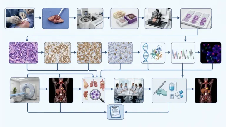
From Diagnosis to Recovery: How Does the Process Proceed?
The diagnosis and treatment process for vaginal cancer progresses in stages depending on the patient’s clinical condition:
- Initial Examination and Evaluation: A detailed medical history is taken from patients presenting with suspected vaginal cancer. Menopausal status, menstrual cycle, history of abnormal bleeding and discharge, postcoital bleeding, pelvic pain, and urinary/bowel symptoms are assessed. A history of previous cervical or vulvar cancer, HPV infection, smoking, DES exposure, and HIV status is assessed. During the physical examination, the vaginal wall, cervix, external genitalia, and inguinal lymph nodes are examined in detail.
- Tests and Imaging: The entire vaginal wall is visualized via speculum examination and colposcopy; Lugol’s solution highlights dysplastic areas. A biopsy is taken from any suspicious lesion; histopathological diagnosis and HPV status are evaluated. Pelvic MRI is the gold standard for assessing local tumor spread. Chest-abdominal-pelvic CT and PET-CT are performed to screen for distant metastases. Cystoscopy and proctoscopy evaluate bladder and rectal involvement in advanced stages.
- Treatment Planning: Findings are reviewed by a multidisciplinary gynecologic oncology board (gynecologic oncologist, radiation oncologist, medical oncologist, radiologist, and pathologist). The treatment strategy is determined based on the FIGO stage: Local treatment for Stage 0; in Stage I, surgery or brachytherapy; in Stages II–IVa, chemoradiotherapy + brachytherapy; and in Stage IVb, systemic chemotherapy is planned. The location of the tumor within the vagina—in which of the three segments—directly influences the surgical plan.
- Surgical Procedure and Hospital Stay: Vaginal cancer surgery is performed under general anesthesia. A radical hysterovaginectomy takes 2–4 hours; a vaginectomy may take less time. The hospital stay is 3–7 days, depending on the extent of the surgery. Postoperative pain management, wound care, urinary catheter monitoring, and early mobilization are closely monitored. The decision regarding adjuvant therapy is made based on pathology results.
- Recovery After Discharge: Full recovery occurs within 4–8 weeks. The use of a vaginal dilator is recommended due to the risk of vaginal stenosis following radiation therapy. Lymphedema management (compression stockings, physical therapy) may be necessary. Sexual function and psychosocial support should be assessed. As part of the follow-up plan, gynecological examinations, vaginal cuff smears, and imaging are recommended every 3 months for the first 2 years and every 6 months for the following 3 years. The risk of recurrence is highest during the first 2 years.
According to Assoc. Prof. Dr. Nilay Şengül, in the follow-up process after vaginal cancer treatment, the management of vaginal stenosis and lymphedema is critical, especially in patients who have undergone radiation therapy; these complications directly affect quality of life and require early intervention.
Frequently Asked Questions
Is Vaginal Cancer Fatal?
The prognosis for vaginal cancer varies depending on the stage. While the 5-year survival rate is 69% for localized cancer (Stage I), this rate drops to 26% for advanced-stage cancer (Stage IV). According to studies, the overall 5-year survival rate ranges from 44% to 63%. Early diagnosis and appropriate treatment significantly improve survival. However, many patients are in advanced stages at the time of diagnosis, which negatively affects the prognosis. Regular follow-up and HPV vaccination are critical for early diagnosis and prevention.
Is Vaginal Cancer Contagious?
Vaginal cancer itself is not contagious. However, HPV (human papillomavirus), the most significant risk factor for vaginal cancer, can be transmitted sexually. HPV infection is very common; however, most infected women do not develop vaginal cancer. The HPV vaccine reduces the risk of both cervical and vaginal cancer. Everyday social contact, using the restroom, or swimming in a pool does not spread vaginal cancer.
Is Vaginal Cancer Dangerous?
Vaginal cancer is a treatable form of cancer if diagnosed in the early stages. However, the prognosis worsens as diagnosis is delayed. In localized forms, the 5-year survival rate is69, while in advanced stages, this rate drops to 26%. The degree of risk varies depending on the stage, histological type, and response to treatment. Squamous cell carcinoma generally has a better prognosis than adenocarcinoma. Early diagnosis is possible through regular gynecological examinations and HPV screening.
Vaginal cancer is a rare but serious malignancy in women. Squamous cell carcinoma is the most common histological type and is strongly associated with HPV infection. It may not present symptoms in the early stages; therefore, evaluating symptoms such as abnormal bleeding, discharge, and pelvic pain after menopause is critical. Treatment involves a combination of surgery, radiation therapy, and chemotherapy, depending on the stage. HPV vaccination and regular gynecological screening are vital for both prevention and early diagnosis.
To discuss your personal situation regarding vaginal cancer and have your questions answered, you can consult Assoc. Prof. Dr. Nilay Şengül. To schedule an appointment at her clinic at Memorial Şişli Hospital, please call 444 7 888.
Sources
- PMC/NIH, “Incidence and survival of vulvar and vaginal cancers in a population-based cohort” — https://pmc.ncbi.nlm.nih.gov/articles/PMC12550708/
- NCBI Bookshelf/NIH, “Vaginal Cancer – StatPearls” — https://www.ncbi.nlm.nih.gov/books/NBK559126/
- Mayo Clinic, “Vaginal cancer – Diagnosis and treatment” — https://www.mayoclinic.org/diseases-conditions/vaginal-cancer/diagnosis-treatment/drc-203524
- NCCN Guidelines, Vulvar Cancer (Vaginal Cancer Management Principles) — https://www.nccn.org/professionals/physician_gls/pdf/vulvar.pdf
- ESGO Guidelines, Cancer of the Vagina — https://www.esgo.org/guidelines/guidelines/cancer-of-the-vagina/
- PubMed/NIH, “Human papillomavirus and cervical/vaginal cancer” — https://pubmed.ncbi.nlm.nih.gov/29368388/
- American Cancer Society, “Vaginal Cancer Statistics” — https://www.cancer.org/cancer/types/vaginal-cancer/about/key-statistics.html