📌 What You Need to Know in a Nutshell
- Approximately 15% of renal masses are benign; the most common malignant type is renal cell carcinoma.
- According to the Bosniak classification, Type I–II masses are benign, while Type III–IV masses require surgical evaluation.
- In T1a-stage (<4 cm) kidney cancers, partial nephrectomy achieves a 5-year survival rate of 90–95%.
- Active surveillance is a safe alternative treatment option for patients with T1a-stage disease and those at high surgical risk.
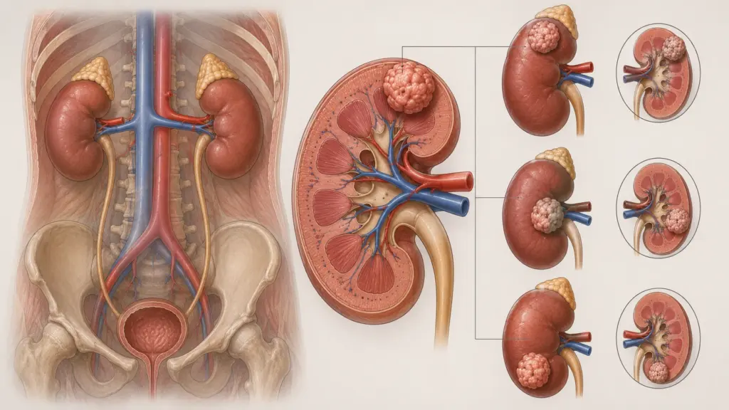
A renal mass refers to cystic (fluid-filled) or solid (tissue-containing) lesions detected in the renal parenchyma. According to the 2024 EAU guidelines, approximately 15% of kidney tumors are benign; the most common malignant types are clear cell, papillary, and chromophobe renal cell carcinomas. Early diagnosis and appropriate treatment are critical for preserving kidney function and improving survival.
What Is a Renal Mass?
A renal mass is a general term that encompasses any cystic or solid lesion developing in kidney tissue and detectable by radiological imaging. These lesions may be benign (oncocytoma, angiomyolipoma, simple cyst) or malignant (renal cell carcinoma, urothelial carcinoma, metastasis). According to the 2024 EAU guidelines, approximately 15% of kidney tumors are benign, and the most common benign types are oncocytoma and angiomyolipoma.
Renal masses may be detected incidentally (on an ultrasound or CT scan performed for another reason) or may cause symptoms such as bloody urine, flank pain, or a palpable mass. With the widespread use of imaging techniques in recent years, the vast majority of renal masses (60–70%) are diagnosed incidentally.
According to Assoc. Prof. Dr. Nilay Şengül, the most critical step in evaluating a renal mass is accurately determining whether the lesion is cystic or solid. According to the protocol we follow at Memorial Şişli Hospital, the Bosniak classification is performed using contrast-enhanced CT or MRI in every patient with a suspected renal mass. This approach reduces unnecessary surgical interventions while preventing the missed diagnosis of malignant lesions.
Types of Renal Masses and Classification
Renal masses are evaluated using the Bosniak classification based on their radiological characteristics. This classification is the standard for determining the risk of malignancy in cystic lesions.
Bosniak Classification (EAU 2024 Update)
| Type | Radiological Characteristics | Risk of Malignancy | Treatment |
|---|---|---|---|
| I | Simple cyst; thin, smooth wall, homogeneous fluid, no septa | 0% | No follow-up required |
| II | Minimal septa, fine calcifications, homogeneous hyperattenuation <3 cm | 0% | No follow-up required |
| IIF | Numerous minimal septa, minimal wall thickening | 5–10% | Follow-up for up to 5 years |
| III | Thick, irregular wall/septa, contrast-enhancing nodule | >50% | Surgery or active surveillance |
| IV | Contrast-enhancing solid component, nodule with well-defined margins | >90% | Surgery |
According to the 2024 EAU guidelines, Bosniak type III cysts should be managed like localized RCC, or active surveillance should be recommended. Type IV cysts, on the other hand, require surgical treatment similar to that for localized RCC. In the 2024 EAU update, the follow-up period for Type IIF lesions has been limited to 5 years.
Benign Renal Lesions
Oncosarcoma: This is the most common benign kidney tumor. According to the 2024 EAU guidelines, active surveillance may be considered as an alternative to surgery or ablation in patients diagnosed with oncosarcoma via biopsy. However, a definitive diagnosis of oncocytoma cannot be made by needle biopsy, as chromophobe RCC may contain areas that closely resemble oncocytoma.
Angiomyolipoma (AML): A benign tumor composed of fat, muscle, and vascular tissue. It is more common in women. According to the 2024 EAU guidelines, selective arterial embolization or nephron-sparing surgery is recommended for large tumors, pregnant women, patients with limited access to emergency medical care, and those with recurrent bleeding or pain. Everolimus may be used in cases of AML where surgical resection is not possible.
Malignant Renal Tumors
Clear-cell renal cell carcinoma (ccRCC): Accounts for 70–75% of all RCCs. According to the 2024 EAU guidelines, partial nephrectomy (PN) is the gold standard for localized ccRCC. Robotic and laparoscopic PN offer the advantages of a shorter hospital stay and less bleeding compared to open PN.
Papillary RCC: Accounts for 10–15% of all RCCs. Type 1 papillary RCC has a better prognosis. In metastatic papillary RCC, casentinib has extended progression-free survival compared to sunitinib.
Chromophobe RCC: Accounts for 5% of all RCCs. It generally has a better prognosis. Differentiation from oncocytoma can be challenging.
Right Renal Mass and Left Renal Mass
Renal masses can occur with equal frequency in the right or left kidney. However, there are some differences in terms of mass location, surgical approach, and anatomical relationships.
Right Renal Mass
The right kidney is located immediately below the liver. According to the 2024 EAU guidelines, liver mobilization may be required during laparoscopic or robotic surgery for upper-pole masses of the right kidney. The right renal vein drains into the inferior vena cava (IVC) at a short distance; therefore, in cases involving venous tumor thrombi, surgery on the right side may be technically easier. Masses in the right kidney may present with symptoms earlier due to pressure on the liver.
Left Renal Mass
The left kidney is adjacent to the spleen and the tail of the pancreas. According to the 2024 EAU guidelines, preserving the spleen and pancreas during partial nephrectomy of the left kidney requires more careful planning. The left renal vein drains into the inferior vena cava (IVC) over a longer distance; this influences surgical planning regarding the removal of venous thrombi. Due to the proximity of the pancreas and spleen, the value of MRI imaging in the preoperative evaluation of left kidney masses is more pronounced.
| Characteristics | Right Kidney | Left Kidney |
|---|---|---|
| Adjacent Organs | Liver, right colon | Spleen, tail of the pancreas, left colon |
| Length of the renal vein | Short (2–4 cm) | Long (6–10 cm) |
| Surgery for venous thrombus | Technically easier | Requires dissection of a longer vein |
| Surgical approach | Transperitoneal is preferred | Transperitoneal or retroperitoneal |
Treatment of a Renal Mass
Treatment of a renal mass is tailored to the size, stage, and histology of the lesion, as well as the patient’s overall condition. According to the 2024 EAU guidelines, treatment options include surgery, ablative therapies, active surveillance, and systemic therapy.
Surgical Treatment
Partial nephrectomy (PN): According to the 2024 EAU guidelines, nephron-sparing surgery (NSS) should be preferred over radical nephrectomy (RN) in localized RCC. PN is strongly recommended for T1a (<4 cm) tumors. For T2 tumors and in patients with a solitary kidney or chronic kidney disease, PN may be considered if technically feasible. Robotic and laparoscopic PN result in shorter hospital stays and less bleeding compared to open PN.
Radical nephrectomy (RN): It is recommended for T2 tumors and in cases where PN is not technically feasible (locally advanced stage, unsuitable tumor location). Laparoscopic RN is associated with lower morbidity compared to open RN. According to the 2024 EAU guidelines, RN should not be performed in patients with T1 tumors if PN is feasible.
Lymph node dissection: According to the 2024 EAU guidelines, the survival benefit of extended lymph node dissection in localized disease has not been proven. However, clinically enlarged lymph nodes should be removed for staging and prognosis.
Ablative Therapies
Radiofrequency ablation (RFA) and cryotherapy may be used in selected patients with T1a-stage renal masses. According to the 2024 EAU guidelines, these treatments may be considered particularly in elderly patients and those with comorbidities who are at high surgical risk. However, long-term oncological outcomes are less well-established compared to surgery.
Active Surveillance
Active surveillance is a safe alternative for patients with stage T1a disease and those at high surgical risk. According to the 2024 EAU guidelines, active surveillance is an acceptable alternative to surgery or ablation in patients diagnosed with oncocytoma or other oncocytic renal tumors based on biopsy findings. The follow-up protocol is determined based on tumor size and growth rate.
Systemic Therapies
In metastatic renal cell carcinoma (mRCC), immune checkpoint inhibitor (ICI) combinations are recommended as first-line therapy. According to the 2024 EAU guidelines:
- IMDC Favorable Risk: Nivolumab + casentinib, pembrolizumab + axitinib, pembrolizumab + lenvatinib, nivolumab + ipilimumab
- IMDC Intermediate/Poor Risk: Nivolumab + casentinib, pembrolizumab + axitinib, pembrolizumab + lenvatinib, nivolumab + ipilimumab
According to the CheckMate 025 study, nivolumab provided superior overall survival compared to everolimus (5-year OS: 26% vs. 18%; HR: 0.73). The pembrolizumab + axitinib combination significantly prolonged survival compared to sunitinib.
[VISUAL SUGGESTION: Renal mass treatment algorithm diagram — showing surgical, ablation, active surveillance, and systemic treatment options based on stage and risk status — Alt text: "Stage-based renal mass treatment algorithm decision tree diagram"]
Clinical Assessment by Assoc. Prof. Dr. Nilay Şengül
In my clinical practice, I frequently observe the following: Patients with incidentally detected small renal masses (<4 cm) often express concerns about overtreatment. In the cases we evaluated at Memorial Şişli Hospital, in 30–40% of patients diagnosed at stage T1a, both cancer control and kidney function can be preserved through active surveillance or partial nephrectomy.
According to Assoc. Prof. Dr. Nilay Şengül, the most significant paradigm shift in the treatment of renal masses is the transition from the approach of “removing every renal mass via radical nephrectomy” to “nephron-sparing surgery and personalized treatment.” Particularly in stage T1a, survival rates with robotic partial nephrectomy are equivalent to those with radical nephrectomy; however, kidney function is preserved significantly better. This approach reduces the future risk of chronic kidney disease and cardiovascular complications.
In metastatic RCC, combinations of immunotherapy and targeted therapy have fundamentally transformed survival over the past 5 years. Particularly in patients with intermediate and poor IMDC risk, median survival can reach 4–5 years with combinations of nivolumab + ipilimumab or pembrolizumab + axitinib.
From Diagnosis to Recovery: How Does the Process Proceed?
- Initial Examination and Evaluation: The patient’s symptoms (bloody urine, flank pain, weight loss), family history, and general health status are assessed. During the physical examination, the abdomen is palpated and lymph node regions are evaluated. Assoc. Prof. Dr. Şengül emphasizes that in her clinical practice, evaluating the patient’s kidney function (creatinine, GFR) and comorbidities (hypertension, diabetes) during the initial consultation directly influences the treatment plan.
- Diagnostic Tests:
- Contrast-enhanced CT (abdomen + chest): The gold standard for diagnosing and staging renal masses; it assesses tumor size, venous invasion, and lymph node and lung metastases
- MRI: Preferred for evaluating venous involvement, or if radiation exposure needs to be avoided or there are contraindications to CT contrast
- Contrast-enhanced ultrasound: Aids in the characterization of small renal masses
- Biopsy: Mandatory in patients scheduled for ablative therapy or systemic treatment; core biopsy using the coaxial technique is recommended
- Staging and Risk Assessment: AJCC 8th edition TNM staging is applied. T1a: <4 cm, T1b: 4–7 cm, T2a: 7–10 cm, T2b: >10 cm, T3: venous/perinephric invasion, T4: extension beyond Gerota’s fascia. In patients with metastatic disease, risk stratification is performed using the IMDC (International Metastatic RCC Database Consortium) criteria.
- Treatment Approach:
- T1a, low risk: Partial nephrectomy or active surveillance (depending on the patient)
- T1b–T2, technically feasible: Partial nephrectomy
- T2+, not technically feasible: Radical nephrectomy
- Metastatic: Systemic therapy (combination of ICI and TKI)
- Hospital Stay and Discharge: The hospital stay is 1–3 days for robotic or laparoscopic partial nephrectomy and 2–4 days for radical nephrectomy. For open surgery, this duration may extend to 4–7 days. Patients are provided with information regarding pain management, wound care, and activity restrictions following discharge.
- Follow-up and Monitoring: According to the 2024 EAU guidelines, the follow-up schedule is determined based on the risk profile. Low risk (Leibovich 0–2): Ultrasound/CT every 6 months, then annually after 5 years. High risk (Leibovich ≥6): CT scan every 3 months for 2 years; every 6 months for 3 years; annually for 5 years. Functional follow-up (creatinine, GFR, cardiovascular assessment) continues for life.
Frequently Asked Questions
What is a renal mass?
A renal mass refers to cystic (fluid-filled) or solid (tissue-containing) lesions that develop in kidney tissue. These lesions can be benign (non-cancerous) or malignant (cancerous). According to the 2024 EAU guidelines, approximately 15% of kidney tumors are benign. The most common benign types are oncocytoma and angiomyolipoma; the most common malignant type is clear cell renal cell carcinoma.
Is a kidney mass always cancer?
No. Approximately 15% of kidney masses are benign; oncocytomas, angiomyolipomas, and simple cysts fall into this category. However, the vast majority of masses are malignant. The Bosniak classification is used to determine the risk of malignancy in cystic masses. Type I and II cysts are benign, while Type III and IV cysts require surgical evaluation. As for solid masses, 70–80% are malignant.
What are the symptoms of a kidney mass?
Small renal masses (<4 cm) are usually asymptomatic and are detected incidentally. Symptoms include bloody urine (hematuria), flank pain (lower back pain), a palpable mass, weight loss, fatigue, and night sweats. The classic triad (hematuria, pain, mass) is now rare, as most masses are detected by imaging in the early stages.
How is a renal mass diagnosed?
The diagnosis is made using contrast-enhanced CT (abdomen and chest). CT evaluates tumor size, venous invasion, and lymph node and lung metastases. MRI is preferred for a detailed assessment of venous involvement or if the patient is allergic to CT contrast. A biopsy is mandatory in patients scheduled for ablative therapy or systemic treatment. According to the 2024 EAU guidelines, the core biopsy technique should be preferred over fine-needle aspiration (FNA).
Partial nephrectomy or radical nephrectomy?
According to the 2024 EAU guidelines, partial nephrectomy (PN) should be preferred over radical nephrectomy (RN) in localized renal cell carcinoma. PN ensures cancer control while preserving kidney function. PN is strongly recommended for T1a (<4 cm) tumors. PN may be considered for T2 tumors and in patients with a solitary kidney. However, if PN is not technically feasible due to tumor location or advanced stage, RN is performed.
Is robotic surgery advantageous for renal masses?
According to the 2024 EAU guidelines, robotic and laparoscopic partial nephrectomy offer the advantages of a shorter hospital stay and less bleeding compared to open surgery. However, long-term oncological outcomes (survival, recurrence) are equivalent to those of open surgery. Robotic surgery may facilitate the preservation of surgical margins, particularly in complex tumors (T1b, hilar location). The most appropriate surgical approach for each patient is determined based on tumor characteristics and the surgeon’s experience.
Is active surveillance safe?
Yes, it is safe for selected patients. According to the 2024 EAU guidelines, active surveillance is an acceptable alternative for patients with stage T1a and those at high surgical risk, as well as for patients diagnosed with oncocytoma via biopsy. If, during follow-up, the tumor growth rate exceeds 0.5 cm per year or the total size reaches >4 cm, treatment is initiated. Active surveillance is particularly recommended for elderly patients and those with serious comorbidities.
Can a metastatic renal mass be treated?
Yes. In metastatic renal cell carcinoma, combinations of immune checkpoint inhibitors (ICIs) and tyrosine kinase inhibitors (TKIs) are used as first-line therapy. According to the 2024 EAU guidelines, the combinations of nivolumab + casentinib, pembrolizumab + axitinib, and pembrolizumab + lenvatinib are recommended. With these treatments, median survival can reach 4–5 years. For second-line treatment, options such as casentinib or nivolumab (in ICI-naive patients) are considered.
Renal tumors are a group of diseases for which high survival rates can be achieved through early diagnosis and personalized treatment. The Bosniak classification is the gold standard for managing cystic lesions, while nephron-sparing surgery is the gold standard for localized malignant tumors. In stage T1a, partial nephrectomy or active surveillance ensures both cancer control and the preservation of kidney function. In metastatic disease, combination immunotherapies have significantly improved survival.
To discuss your specific situation regarding a renal mass and have your questions answered, you can consult with Assoc. Prof. Dr. Nilay Şengül. To schedule an appointment at her clinic at Memorial Şişli Hospital, please call 444 7 888.
Sources
- EAU Guidelines on Renal Cell Carcinoma 2024, European Association of Urology — https://d56bochluxqnz.cloudfront.net/documents/full-guideline/EAU-Guidelines-on-Renal-Cell-Carcinoma-2024.pdf
- Medscape, "Renal Cell Carcinoma: EAU 2025 Guideline Summary" — https://reference.medscape.com/cc2/p10/eau-guideline-renal-cell-carcinoma-2024a1000im7
- EAU Pocket Guidelines on Renal Cell Carcinoma 2024 — https://d56bochluxqnz.cloudfront.net/documents/pocket-guidelines/EAU-Pocket-on-Renal-Cell-Carcinoma-2024.pdf
- CheckMate 025 Trial: Nivolumab versus Everolimus in Advanced Renal-Cell Carcinoma, N Engl J Med. 2015;373(19):1803-1813.
- French AFU Cancer Committee Guidelines Update 2022–2024: Management of Kidney Cancer — https://www.urofrance.org/recommandation/french-afu-cancer-committee-guidelines-update-2022-2024-management-of-kidney-cancer/
- EAU Uroweb, Renal Cell Carcinoma Guidelines Summary of Changes 2024 — https://uroweb.org/guidelines/renal-cell-carcinoma/summary-of-changes/2024