📌 What You Need to Know in a Nutshell
- Breast cancer is the most common cancer among women; 288,494 new cases were reported in the U.S. in 2023, and the 5-year overall survival rate is 91%.
- In the early (localized) stage, the 5-year survival rate reaches 99%; in the metastatic stage, this rate drops to 31%; 66% of cases are diagnosed at the localized stage.
- The most common symptom is a painless lump; nipple discharge, skin changes, and underarm swelling are also important signs.
- Treatment is personalized based on stage and biological subtype; combinations of surgery, chemotherapy, radiation therapy, hormone therapy, targeted therapy, and immunotherapy are used.
Breast cancer
is a malignancy originating in breast tissue and is the most common cancer among women. This disease, which has a high treatment success rate when diagnosed early, can be significantly controlled through modern screening programs, advanced surgical techniques, targeted therapies, and immunotherapy. However, thousands of women are diagnosed with metastatic breast cancer each year, and this directly affects treatment outcomes.
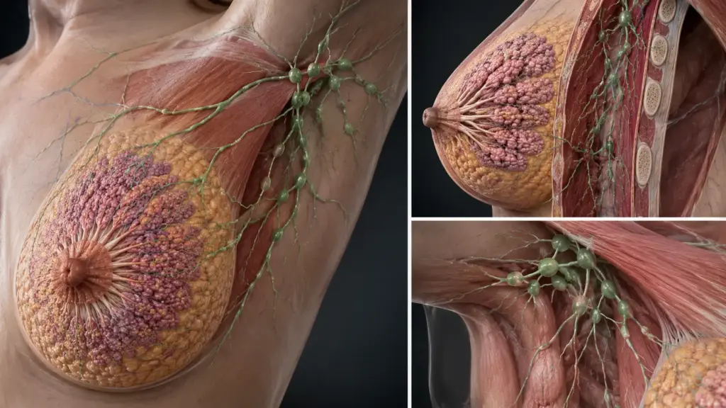
[VISUAL SUGGESTION: Breast anatomy and lymphatic drainage diagram — an anatomical diagram showing the breast lobules, ducts, lymph nodes, and axillary region — Alt text: “Breast anatomy and lymphatic drainage diagram — lobules, ducts, and lymph nodes”]
What Is Breast Cancer?
Breast cancer
is a malignant tumor that develops as a result of the uncontrolled proliferation of cells in breast tissue. Breast tissue consists of lobules (milk-producing glands) and ducts (tubes that carry milk to the nipple). Cancer can originate in any of these structures; it most commonly develops from the epithelial cells lining the inner surface of the ducts (ductal carcinoma).
Histologically, breast cancer is divided into two main groups: invasive ductal carcinoma (IDC, 70–80%) and invasive lobular carcinoma (ILC, 10–15%). Less common subtypes include mucinous, tubular, medullary, and metaplastic carcinomas. Biologically, four subtypes have been identified: luminal A, luminal B, HER2-positive, and triple-negative; these subtypes directly determine treatment selection.
According to CDC U.S. Cancer Statistics data, 288,494 women in the United States were newly diagnosed with breast cancer in 2023, and 43,402 women died from the disease in 2024. Approximately 66% of cases are diagnosed at the localized (local) stage, 25% at the regional (lymph node involvement) stage, and 6% at the distant (metastatic) stage.
According to Assoc. Prof. Dr. Nilay Şengül, the most critical factor in breast cancer diagnosis is early detection; because while the 5-year survival rate reaches 99% at the localized stage, this rate drops to 31% at the metastatic stage. For this reason, regular breast self-exams and mammography screenings are of life-saving importance.
What Are the Symptoms of Breast Cancer?
The most common symptom of breast cancer is a painless, firm lump with irregular edges. However, not every lump is cancer; benign lesions such as fibroadenomas and cysts can also form lumps. Other important symptoms include:
- Nipple discharge: Discharge—especially if it is one-sided, bloody, or clear—is a cause for concern.
- Nipple retraction or change in direction: This occurs when the tumor pulls on the ligaments attached to the nipple.
- Skin changes: Orange peel appearance (peau d’orange), redness, ulceration, or thickening.
- Swelling in the armpit (axillary): A sign of lymph node involvement.
- Changes in breast shape or size: Unilateral enlargement, shrinkage, or asymmetry.
- Persistent pain: Cancer is generally painless; however, pain may develop in advanced stages.
Assoc. Prof. Dr. Şengül emphasizes that in her clinical practice, approximately 50% of patients diagnosed with breast cancer first noticed a lump during a self-examination. For this reason, it is recommended that every woman perform a monthly breast self-exam and undergo an annual mammogram starting at age 40. Symptoms such as nipple discharge or skin changes may occur even without a lump and must be evaluated.
| Symptoms | Frequency | Severity |
|---|---|---|
| Painless lump | Most common | Very high — every mass should be evaluated |
| Nipple discharge | Moderate | High — especially bloody discharge |
| Skin changes | Moderate | High — orange peel appearance |
| Swelling under the arms | Moderate | High — sign of lymph node involvement |
| Nipple retraction | Moderate | High — the tumor is pulling on the ligaments |
What Causes Breast Cancer? Risk Factors
Hormonal, genetic, and environmental factors all play a role in the development of breast cancer. Although the exact cause is unknown, the following factors increase the risk:
- Gender and age: It is much more common in women than in men; the median age at diagnosis is 63. However, it can also occur in women under 50.
- Family history and genetic mutations: BRCA1 and BRCA2 mutations increase the risk of breast cancer to 60–80%. Mutations in other genes, such as TP53, PTEN, and PALB2, also contribute to an increased risk.
- Hormonal factors: Early menarche, late menopause, nulliparity (never having given birth), having the first child after age 30, and hormone replacement therapy increase the risk.
- History of breast cancer: A woman who has had breast cancer in one breast has an increased risk of developing cancer in the other breast.
- Breast density: Dense breast tissue both increases the risk of cancer and makes detection on mammograms more difficult.
- Ionizing radiation: Radiation therapy to the breast area (e.g., treatment for Hodgkin lymphoma) increases the risk.
- Alcohol and obesity: Excessive alcohol consumption and obesity (especially during the postmenopausal period) increase the risk.
- Physical inactivity: Regular exercise reduces the risk.
According to 2025 data from the American Cancer Society, the lifetime risk of developing breast cancer in women is 13.1% (approximately 1 in 8). This risk is much higher in carriers of BRCA1/2 mutations. Protective factors include breastfeeding, physical activity, a healthy weight, and limiting alcohol consumption.
How Is Breast Cancer Diagnosed? The Diagnostic Process
Breast cancer is diagnosed through clinical examination, imaging, and biopsy. Screening programs enable the detection of asymptomatic lumps in the early stages.
Screening
The U.S. Preventive Services Task Force (USPSTF) recommends mammography screening every two years for women aged 40–74. For women with dense breast tissue, breast ultrasound or breast MRI may also be considered. In Turkey, free mammography screening is offered to women aged 40–69 through KETEM centers.
Diagnostic Process
- Clinical examination: Palpation of the breast and axilla, and evaluation of nipple discharge are performed.
- Imaging: Diagnostic mammography, breast ultrasound, and, if necessary, breast MRI are ordered. The degree of suspicion is determined using the BI-RADS scoring system.
- Biopsy: A fine-needle aspiration biopsy (FNAB), tru-cut biopsy, or vacuum-assisted biopsy is performed on the suspicious lesion. This is the gold standard for diagnosis.
- Pathological evaluation: Tumor type, grade, hormone receptor status (ER, PR), HER2 status, and Ki-67 proliferation index are assessed.
- Staging: Breast MRI, CT (thorax-abdomen-pelvis), bone scan, and PET-CT are used to investigate for distant metastases.
According to Assoc. Prof. Dr. Nilay Şengül, the most significant cause of diagnostic delay in breast cancer is patients viewing a lump as a “temporary change” or “not important because it is painless.” On the contrary, a painless lump is the most important sign of cancer; therefore, any lump that persists for more than 2 weeks must be evaluated.
Stages of Breast Cancer
Breast cancer staging is performed according to the 8th edition of the AJCC (American Joint Committee on Cancer) guidelines. The stage is determined based on tumor size (T), lymph node involvement (N), and the presence of distant metastases (M). The stage directly influences the treatment strategy and prognosis.
| Stage | Definition | 5-Year Survival Rate |
|---|---|---|
| 0 (Carcinoma in situ) | Ductal carcinoma in situ (DCIS) | >99% |
| IA | Tumor ≤2 cm, negative lymph nodes | 99% |
| IB | Tumor ≤2 cm, micrometastases (0.2–2 mm) | 98% |
| IIA | Tumor 2–5 cm or ≤2 cm with 1–3 positive lymph nodes | 93 |
| IIB | Tumor >5 cm or 2–5 cm with 1–3 positive lymph nodes | 85 |
| IIIA | 4–9 positive lymph nodes or chest wall involvement | 72% |
| IIIB | Chest wall or skin involvement, 10 or more lymph nodes | 50–60 |
| IIIC | Supraclavicular or internal mammary lymph node | 40–50 |
| IV (Metastatic) | Distant metastasis (bone, liver, lung, brain) | 31 |
According to CDC U.S. Cancer Statistics data, the stage distribution for breast cancer is as follows: Localized stage 66%, regional stage 25%, distant (metastatic) stage 6%. The 5-year survival rate is 99% for the localized stage, 86% for the regional stage, and 31% for the distant stage. The overall 5-year survival rate is 91% (2016–2022 data).
According to 2025 data from the American Cancer Society, breast cancer mortality has decreased by 44% since its peak in 1989; two-thirds of this progress is attributed to advances in treatment, and one-third to early detection through screening. However, the rate of decline has slowed in recent years (2% per year in the 2000s, 1% per year since 2010).
Breast Cancer Treatment
Breast cancer treatment is personalized based on the stage, hormone receptor status (ER/PR), HER2 status, genetic profile, and the patient’s overall condition. A multidisciplinary approach (surgery, medical oncology, radiation oncology, pathology, and radiology) is standard.
Surgical Treatment
Breast-conserving surgery (BCS):
The tumor is removed along with a margin of healthy surrounding tissue (lumpectomy, segmental mastectomy). BCS provides survival rates equivalent to those of mastectomy; however, it must be performed in conjunction with radiation therapy. Patients are candidates for BCS if the tumor is ≤5 cm, solitary, and proportional to breast size.
Mastectomy:
This involves the removal of all breast tissue. Options include modified radical mastectomy (breast plus axillary lymph nodes), subcutaneous mastectomy (with preservation of the nipple), and skin-sparing mastectomy (combined with reconstruction). Prophylactic mastectomy reduces the risk by 90% in BRCA mutation carriers.
Sentinel lymph node biopsy (SLNB):
It is the gold standard for assessing axillary lymph node involvement. If the sentinel node is negative, axillary dissection (removal of all lymph nodes) is not required; this significantly reduces the risk of lymphedema.
Systemic Therapy
Chemotherapy:
Used as adjuvant (post-surgical) or neoadjuvant (pre-surgical) therapy. Doxorubicin, cyclophosphamide, paclitaxel, and docetaxel are commonly used agents. 21st-century oncogenomic research has led to the development of genetic signatures (Oncotype DX, MammaPrint) that predict response to chemotherapy; these tests have demonstrated that chemotherapy is unnecessary in low-risk patients.
Hormone therapy:
Used in ER- and/or PR-positive tumors. Selective estrogen receptor modulators (tamoxifen), aromatase inhibitors (anastrozole, letrozole, exemestane), and estrogen receptor degraders (fulvestrant) are treatment options. Adjuvant hormone therapy is continued for 5–10 years.
HER2-targeted therapy:
Agents such as trastuzumab (Herceptin), pertuzumab (Perjeta), T-DM1 (Kadcyla), and trastuzumab deruxtekan (Enhertu) have significantly improved survival in patients with HER2-positive tumors. The CLEOPATRA trial demonstrated that the combination of trastuzumab and pertuzumab extended median survival to 56.5 months in patients with metastatic HER2-positive breast cancer.
Immunotherapy:
Pembrolizumab has received FDA approval for use in triple-negative breast cancer (TNBC) in both the adjuvant and metastatic settings. The KEYNOTE-522 study showed that the combination of pembrolizumab and chemotherapy increased the neoadjuvant response rate from 51.2% to 64.8%.
CDK4/6 inhibitors:
Palbociclib, ribociclib, and abemaciclib are combined with hormone therapy in HR+ / HER2- metastatic breast cancer; they significantly prolong PFS.
According to Assoc. Prof. Dr. Nilay Şengül, the most critical determinant of treatment success in breast cancer is a “personalized approach”; that is, two patients at the same stage may receive completely different treatments if they belong to different biological subtypes. For example, while a HER2-positive patient receives trastuzumab, a triple-negative patient receives immunotherapy and chemotherapy. For this reason, comprehensive pathological and molecular analysis is mandatory for every patient.
Breast Cancer Surgery: What to Expect?
Breast cancer surgery is planned with a scope that varies depending on the tumor stage, location, and the patient’s preference. The primary goal is the complete removal of the tumor and the assessment of lymph node status.
Preoperative:
Blood tests, an EKG, a chest X-ray, and an anesthesia consultation are performed prior to hospital admission. Anticoagulant medications are discontinued before surgery. A breast MRI helps assess the extent of tumor spread and whether the cancer is multifocal.
Surgical procedure: The procedure
is performed under general anesthesia. Breast-conserving surgery (BCS) takes 1–2 hours, while a mastectomy takes 2–3 hours. A sentinel lymph node biopsy is performed using radiocolloid and blue dye for mapping. Reconstruction (using an implant or autologous tissue) can be performed during the same session or in a second stage, in conjunction with a skin-sparing mastectomy.
Postoperative care:
The hospital stay is 1 day for BCS and 1–3 days for mastectomy. Drains may remain in place for 1–2 weeks. Pain management, wound care, and early mobilization are priorities. In patients who undergo lymph node dissection, the risk of lymphedema (swelling in the arm and hand) ranges from 10% to 30%; in such cases, physical therapy and compression bandages are recommended.
Assoc. Prof. Dr. Şengül notes that the most common issue she encounters in her clinical practice following breast cancer surgery is “patients struggling to make a decision about reconstruction.” Reconstruction not only restores physical appearance but also supports psychological recovery; however, this decision should be left entirely to the patient’s preference. If breast-conserving surgery is an option in the early stages, this should be explained to the patient in detail.
Clinical Assessment by Assoc. Prof. Dr. Nilay Şengül
In my clinical practice, I frequently observe the following: A significant proportion of patients diagnosed with breast cancer experience fear and anxiety at the time of diagnosis; however, thanks to modern treatment options, excellent outcomes can be achieved, particularly in the early stages.
According to Assoc. Prof. Dr. Nilay Şengül, the most critical factor in breast cancer is “personalized treatment”; that is, two patients at the same stage may receive completely different treatments if they have different biological subtypes. For this reason, comprehensive pathological and molecular analysis (Oncotype DX, MammaPrint, BRCA testing) in every patient is essential for treatment success.
Additionally, the first 5 years are the most critical period for posttreatment follow-up in breast cancer, as most recurrences occur during this time. Regular breast examinations, mammograms, and imaging when necessary enable the early detection of recurrence and timely intervention. Furthermore, managing bone health and menopause symptoms in patients receiving hormone therapy is important for maintaining quality of life.
From Diagnosis to Recovery: How Does the Process Proceed?
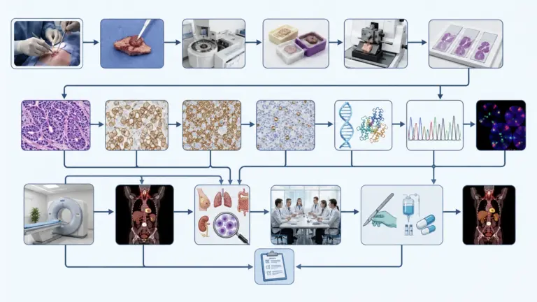
- Initial examination and medical history: During the initial gynecological or general surgical examination, the presence of a lump, discharge, pain, and family history are thoroughly assessed. The presence of breast or ovarian cancer in the family history is recorded. A clinical breast exam and axillary palpation are performed.
- Tests and imaging: Mammography, breast ultrasound, and, if necessary, breast MRI are ordered. A tru-cut or vacuum-assisted biopsy is taken from the suspicious lesion. Once the diagnosis is confirmed, staging is performed using CT, bone scan, and PET-CT.
- Treatment planning: The stage, biological subtype, and treatment strategy are determined during a multidisciplinary tumor board meeting (surgeon, medical oncologist, radiation oncologist, pathologist, radiologist). ER/PR, HER2, Ki-67, and genetic test results are evaluated.
- Surgical procedure: A breast-conserving surgery (BCS) or mastectomy is performed under general anesthesia. A sentinel lymph node biopsy and, if necessary, axillary dissection are performed. Skin-sparing mastectomy and reconstruction can be performed during the same session or at a later date.
- Hospital Stay and Discharge: Patients stay in the hospital for 1 day after BCS and 1–3 days after mastectomy. Discharge occurs after drains are removed. Patients are instructed on wound care, pain management, and arm exercises.
- Recovery and Follow-Up: Adjuvant therapy (chemotherapy, hormone therapy, radiation therapy, targeted therapy) is initiated within 2–4 weeks after surgery. Follow-up is recommended every 3–6 months for the first 2 years, every 6–12 months for years 3–5, and annually thereafter. Follow-up includes a breast examination, mammogram, axillary palpation, and imaging if necessary. Physical therapy and compression bandages are recommended for patients who develop lymphedema.
According to Assoc. Prof. Dr. Nilay Şengül, the first 5 years are the most critical period in post-treatment follow-up for breast cancer, as most recurrences occur during this time. Regular mammograms and clinical examinations enable the early detection of recurrences and prompt intervention. Additionally, managing bone health and menopause symptoms in patients receiving hormone therapy is important for maintaining quality of life.
Frequently Asked Questions
Does breast cancer cause pain?
Early-stage breast cancer is generally painless; the most common symptom is a painless lump. Pain usually occurs in advanced stages as a result of the tumor spreading to the chest wall or adjacent structures. However, breast pain is most often due to benign causes (cysts, fibroadenomas, premenstrual symptoms); nevertheless, evaluation is necessary in suspicious cases.
Is breast cancer genetic?
About 5–10% of breast cancer cases are linked to hereditary gene mutations. BRCA1 and BRCA2 mutations are the most common causes; the lifetime risk for carriers reaches 60–80%. Mutations in other genes, such as TP53, PTEN, PALB2, and CHEK2, can also increase the risk. Genetic counseling and testing are recommended for patients with a family history of the disease.
What are the types of breast cancer?
Histologically, there are invasive ductal carcinoma (70–80%), invasive lobular carcinoma (10–15%), and rare subtypes (mucinous, tubular, medullary, metaplastic). Biologically, four subtypes have been defined: luminal A (ER+/PR+/HER2-/low Ki-67), luminal B (ER+/PR+/HER2- or HER2+/high Ki-67), HER2-positive, and triple-negative (ER-/PR-/HER2-). These subtypes directly determine treatment selection.
Can men get breast cancer?
Yes, men can develop breast cancer; however, it is very rare (1% of all breast cancer cases). Invasive ductal carcinoma is the most common type in men. Risk factors include: BRCA2 mutation, Klinefelter syndrome, family history, radiation exposure, liver disease (impaired estrogen metabolism), and obesity. Diagnosis is often delayed in men; therefore, the prognosis is generally poorer.
What helps with breast cancer?
Breast cancer treatment is personalized based on the stage and biological subtype: surgery (breast-conserving surgery or mastectomy), chemotherapy, radiation therapy, hormone therapy, HER2-targeted therapy (trastuzumab, pertuzumab), and immunotherapy (pembrolizumab, for triple-negative breast cancer). “Remedies” outside of standard treatment (herbal products, alternative therapies) lack scientific support and cannot replace standard treatment.
How long does breast cancer surgery take?
Breast-conserving surgery (BCS) takes 1–2 hours, while a mastectomy takes 2–3 hours. If a sentinel lymph node biopsy is performed, the procedure is extended by 30–60 minutes. If skin-sparing mastectomy and reconstruction (using an implant or autologous tissue) are performed during the same session, the procedure can take up to 4–6 hours. The duration of the surgery depends on the surgeon’s experience and the complexity of the tumor.
Is breast cancer fatal?
The prognosis for breast cancer varies depending on the stage. In the early (localized) stage, the 5-year survival rate is 99%, and curative treatment is possible. The survival rate is 86% in the regional stage and 31% in the metastatic stage. The overall 5-year survival rate is 91%. According to data from the American Cancer Society, mortality has decreased by 44% since 1989; this progress is due to advances in treatment and early diagnosis.
Which department should you visit for breast cancer?
The departments of general surgery (breast surgery), medical oncology, radiation oncology, and gynecologic oncology are involved in the diagnosis and treatment of breast cancer. The initial consultation is usually with a general surgeon or a gynecologist. After diagnosis, a treatment plan is developed during a multidisciplinary tumor board meeting. Treatment at centers specializing in breast cancer (oncology centers) has a positive impact on outcomes.
Which doctor treats breast cancer?
In the diagnosis and treatment of breast cancer, a breast surgeon (general surgeon), medical oncologist, radiation oncologist, pathologist, radiologist, plastic surgeon, and genetic counselor work together. The initial evaluation is usually performed by a general surgeon. Treatment planning is carried out using a multidisciplinary approach; this approach increases treatment success and patient satisfaction.
Breast cancer is the most common cancer in women and has a high treatment success rate when diagnosed early. In the U.S., 288,494 new cases were reported in 2023, and the 5-year overall survival rate reached 91%. While the 5-year survival rate is 99% in the early (localized) stage, it drops to 31% in the metastatic stage. Treatment is personalized based on stage and biological subtype (luminal A/B, HER2+, triple-negative); combinations of surgery, chemotherapy, radiation therapy, hormone therapy, targeted therapy, and immunotherapy are used. Regular mammograms and breast self-exams are critical for early diagnosis and saving lives.
To discuss your personal situation regarding breast cancer and have your questions answered, you can consult with Assoc. Prof. Dr. Nilay Şengül. To schedule an appointment at her clinic at Memorial Şişli Hospital, please call 444 7 888
.
Sources
- CDC, U.S. Cancer Statistics, “Female Breast Cancer Stat Bite” — https://www.cdc.gov/united-states-cancer-statistics/publications/breast-cancer-stat-bite.html
- Siegel RL et al., CA: A Cancer Journal for Clinicians, “Cancer Statistics, 2025” — https://pmc.ncbi.nlm.nih.gov/articles/PMC11745215/
- SEER (National Cancer Institute), “Cancer Stat Facts: Female Breast Cancer” — https://seer.cancer.gov/statfacts/html/breast.html
- NCCN, “Breast Cancer, Version 5.2024, NCCN Clinical Practice Guidelines in Oncology” — https://jnccn.org/view/journals/jnccn/22/5/article-p331.xml
- ESMO, “Early Breast Cancer: ESMO Clinical Practice Guideline for Diagnosis, Treatment, and Follow-Up” — https://www.esmo.org/guidelines/guidelines-by-topic/breast-cancer
- USPSTF, “Screening for Breast Cancer” — https://www.uspreventiveservicestaskforce.org/uspstf/recommendation/breast-cancer-screening