📌 What You Need to Know in a Nutshell
- Ewing sarcoma accounts for 10–15% of bone sarcomas; it most commonly occurs in adolescents and young adults aged 10–20.
- While the 5-year survival rate is 75–80% for localized disease, it drops below 30% for metastatic disease.
- The EWSR1-FLI1 chromosomal translocation is detected in 85% of cases; and this fusion gene plays a key role in diagnostic and targeted therapy research.
- The VDC/IE (vincristine/doxorubicin/cyclophosphamide + ifosfamide/etoposide) combination is the standard chemotherapy regimen for localized disease.
Ewing sarcoma is an aggressive, malignant small round-cell tumor originating in bone and soft tissue. According to NIH/NCBI StatPearls data, it accounts for 10–15% of all bone sarcomas and is most commonly seen in adolescents and young adults aged 10–20. According to the DelveInsight 2026 epidemiology report, approximately 1,250 new cases are diagnosed annually in the 7MM region (U.S., EU4, UK, Japan). With early diagnosis and multimodal treatment, survival rates have risen from 20% to over 70% in the last 40 years.
What Is Ewing Sarcoma?
Ewing sarcoma is an aggressive, malignant small-round-cell tumor originating in bone or soft tissue. It was first described by James Ewing in 1921. Classic bone Ewing sarcoma, extraskeletal Ewing sarcoma, Askin tumor (chest wall), and primitive neuroectodermal tumors (PNET) all belong to the same tumor family. These tumors exhibit similar histological and immunohistochemical characteristics and originate from common mesenchymal progenitor cells.
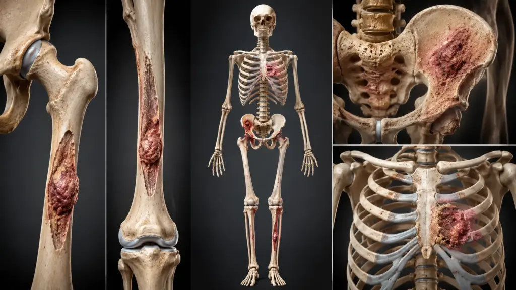
According to NIH/NCBI data, Ewing sarcoma most commonly occurs in the diaphysis and diaphysis-metaphysis regions of long bones such as the femur, tibia, and humerus, as well as in the pelvis and thoracic cage. Extraskeletal forms account for approximately 12% of cases and can develop in virtually any anatomical region. It is 1.4 times more common in males than in females. The highest incidence is reported among individuals of European descent; it is rarer among individuals of African and Asian descent.
According to Assoc. Prof. Dr. Nilay Şengül, the most critical step in diagnosing Ewing sarcoma is ensuring that persistent bone pain in adolescents and young adults is not dismissed as “growing pains.” According to the protocol we follow at Memorial Şişli Hospital, young patients with nighttime pain lasting longer than 2–3 weeks are always evaluated with imaging and biopsy. This approach prevents diagnostic delays and increases treatment success.
What Is the Most Dangerous Sarcoma?
The definition of the “most dangerous” sarcoma varies depending on stage, histological grade, location, and response to treatment. Ewing sarcoma is one of the most serious bone sarcomas due to its aggressive biological behavior and potential for early metastasis. Tumors located in the pelvis and axial skeleton (spine, rib cage), in particular, have a poorer prognosis due to large size, early metastasis, and resistance to chemotherapy.
According to data from Cancer Therapy Advisor, the 5-year survival rate for metastatic Ewing sarcoma is less than 30%. While this rate is 30–40% in patients with isolated lung metastases, it drops to 5–10% in those with bone and bone marrow metastases. There is no standard treatment for recurrent or refractory disease, and survival is below 20%. Therefore, Ewing sarcoma ranks among the most aggressive sarcomas in its untreated or recurrent form.
Other high-risk sarcomas include angiosarcoma (5-year survival rate of 33%), pleomorphic rhabdomyosarcoma, and desmoplastic small round cell tumor. However, due to poses the greatest threat to this age group due to its prevalence in childhood and adolescence.
Symptoms of Ewing Sarcoma
The symptoms of Ewing sarcoma vary depending on the tumor’s location, size, and stage. The most common initial symptom is persistent bone pain that continues even at rest. According to NIH/NCBI data, more than 50% of cases involve nighttime pain.
- Persistent bone pain (especially at night and during rest)
- Swelling and tenderness (in the tumor area)
- Mass (may be palpable, warm, or red)
- Limited mobility (when located near a joint)
- Pathological fracture (due to bone weakening)
- Systemic symptoms: fever, weight loss, fatigue (in metastatic disease)
- Back pain (in pelvic tumors, due to spinal compression)
Ewing Sarcoma Lung Metastasis
The lung is the most common site of distant metastasis for Ewing sarcoma. In 20% of cases, lung or pleural involvement is present at the time of diagnosis. According to NIH/NCBI data, the 5-year overall survival rate is 30–40% in patients with isolated lung metastases; this rate is 5–10% in patients with bone and bone marrow metastases. Symptoms of lung metastases include shortness of breath, cough, chest pain, and hemoptysis. Metastasisectomy (surgical removal) can increase 3-year overall survival to 60% and disease-free survival to 45% in selected patients.
Ewing Sarcoma Genetics
Ewing sarcoma family tumors are characterized by fusion genes resulting from chromosomal translocations. These translocations occur between TET family proteins (EWSR1, FUS) and ETS family transcription factors (FLI1, ERG).
According to NIH/NCBI data, the EWSR1-FLI1 fusion gene results from the t(11;22)(q24;q12) translocation in 85% of cases. In the remaining 10–15%, EWSR1-ERG or other fusion genes are detected as a result of the t(21;12)(22;12) translocation and other rare translocations. These fusion genes encode abnormal transcription factors and disrupt cell proliferation, differentiation, and apoptosis.
The genetic alterations identified in Ewing sarcoma are not hereditary; meaning they do not run in families. No association with environmental risk factors or a history of radiation exposure has been established. The only known risk factors are young age, Caucasian race, and male gender. Molecular cytogenetic analysis is mandatory for diagnosis and serves as the basis for targeted therapy research.
| Genetic Alteration | Prevalence | Clinical Significance |
|---|---|---|
| t(11;22)(q24;q12) — EWSR1-FLI1 | 85% | Gold standard for diagnosis; targeted therapy research |
| t(21;22)(q22;q12) — EWSR1-ERG | 10–15% | May have a more aggressive course |
| Other EWSR1-ETS fusions | <5% | Rare; prognosis variable |
Ewing Sarcoma Stages
Ewing sarcoma staging is based on the TNM system and the presence of metastasis. The most important prognostic factor is the presence of metastasis at the time of diagnosis.
| Stage / Status | Definition | 5-Year Survival Rate |
|---|---|---|
| Localized | Tumor confined to the primary site, no metastasis | 75–80% |
| Metastatic | Metastasis to distant organs present | <%30 |
| Isolated lung metastasis | Lung/pleural involvement only | 30–40% |
| Bone/bone marrow metastasis | Bone or bone marrow involvement | 5–10% |
According to EUROCARE-6 data, the 5-year relative survival rate for Ewing sarcoma in adolescents and young adults (ages 15–39) in Europe is 52%. This rate is lower in children (69%) and older adults. The prognosis worsens with age; it has been reported as 89.8% for ages 1–4, 83.6% for ages 5–9, 75.2% for ages 10–14, and 70.8% for ages 15–19.
Ewing Sarcoma Treatment
Ewing sarcoma treatment requires a multidisciplinary approach. According to the guidelines of the NCCN, ESMO, and GEIS (Spanish Sarcoma Research Group) guidelines, a combination of systemic chemotherapy, surgery, and radiation therapy is administered. Treatment should be planned at an experienced sarcoma center.
Chemotherapy
Chemotherapy is the cornerstone of Ewing sarcoma treatment. According to NCCN guidelines, the VDC/IE (vincristine, doxorubicin, cyclophosphamide + ifosfamide, etoposide) alternating regimen is the standard treatment for localized disease. According to NIH/NCBI data, in the COG AEWS0031 study, VDC/IE administered at 2-week intervals was found to be more effective than at 3-week intervals (without an increase in toxicity).
Induction chemotherapy is planned for 6 cycles (12 weeks). Following local treatment, postoperative chemotherapy may be extended up to 42–48 weeks. In metastatic disease, longer-duration and more intensive chemotherapy regimens may be used. According to ESMO guidelines, local treatment following 3–6 cycles of induction chemotherapy, followed by 6–10 cycles of consolidation chemotherapy, is recommended.
Surgical Treatment
Surgical resection is the preferred method of local control in localized disease. According to ESMO guidelines, wide excision is preferred over radiation therapy. According to NCCN guidelines, wide excision is a Category 1 recommendation. If the surgical margin is positive or very close to the tumor, adjuvant radiation therapy is added.
In metastatic disease, resection of the primary tumor may be considered for palliative purposes or in patients with a limited metastatic burden. According to NIH/NCBI data, an analysis of the SEER database showed that 5-year overall survival and cancer-specific survival were significantly improved in patients with metastatic Ewing sarcoma who underwent local resection. In selected patients with isolated lung metastases, metastasectomycan increase 3-year overall survival to 60%.
Radiotherapy
Radiotherapy is used for local control in patients for whom surgical resection is not feasible or who have positive surgical margins. According to NCCN guidelines, definitive radiation therapy or adjuvant radiation therapy following surgery may be administered. For tumors of the pelvis and axial skeleton, radiation therapy is more commonly preferred due to the difficulty of surgical resection.
Recurrent/Refractory Disease
There is no standard treatment for recurrent/refractory Ewing sarcoma, and the prognosis is poor. According to NIH/NCBI data, 70% of cases relapse within the first 2 years. In patients with local recurrence who are eligible for re-resection, the 5-year survival rate is 30%, whereas in those ineligible for re-resection, this rate is 9%. Participation in clinical trials is the most important option for these patients. High-dose chemotherapy plus autologous stem cell transplant may be considered in selected refractory/relapsed patients.
Ewing Sarcoma Surgery
Ewing sarcoma surgery aims to remove the tumor with wide surgical margins. The surgical technique is determined based on the tumor’s location, size, and relationship to surrounding tissues.
Surgical Techniques
Wide excision: The tumor is removed along with a margin of healthy tissue. This is the preferred surgical approach for Ewing sarcoma. For tumors of the extremities, limb-sparing surgery is performed whenever possible. According to NCCN guidelines, amputation should be considered only in selected cases.
Pelvic tumors: The pelvis is one of the most common sites for Ewing sarcoma. Surgery is technically challenging because the pelvis is adjacent to many vital structures (blood vessels, nerves, bladder, intestines). Wide resection should be planned to minimize functional loss.
Axial skeleton tumors: Tumors of the spine, rib cage (ribs, sternum, clavicle), and sacrum—constitute the most surgically challenging group. These tumors are typically large, metastasize early, and may be resistant to chemotherapy. A combination of surgery and radiation therapy is frequently used.
Postoperative Complications
According to NIH/NCBI data, complications related to surgery or multimodal therapy develop in 25% of Ewing sarcoma treatments. These include delayed wound healing, fibrosis, thrombosis, abscess formation, and implant loosening or fracture. The risk of complications varies depending on the tumor location (pelvis vs. extremity). Assoc. Prof. Dr. Şengül emphasizes that in her clinical practice, wound complications following pelvic Ewing sarcoma surgery are more common than those following extremity surgery.
Clinical Assessment by Assoc. Prof. Dr. Nilay Şengül
In my clinical practice, I frequently observe the following: Adolescent and young adult patients diagnosed with Ewing sarcoma are typically evaluated as having “growing pains” or a sports injury for an average of 2–3 months prior to diagnosis, and this delay leads to disease progression. According to the protocol we follow at Memorial Şişli Hospital, rapid imaging and biopsy are scheduled for young patients with persistent bone pain.
According to Assoc. Prof. Dr. Nilay Şengül, the most significant paradigm shift in Ewing sarcoma treatment is the shift from a “local treatment only” approach to “multimodal systemic treatment.” Over the past 40 years, survival rates for localized disease have risen from 20% to over 70% with VDC/IE chemotherapy. However, there remains a significant unmet need for treatment in metastatic and relapsed/refractory disease. Assoc. Prof. Dr. Şengül notes that new therapies targeting the EWSR1-FLI1 fusion (insulin-like growth factor 1 receptor inhibitors, LSD1 inhibitors, EWS-FLI1-dependent cell death inducers) have yielded promising results in clinical trials. This approach could usher in a new era, particularly in relapsed/refractory disease.
From Diagnosis to Recovery: How Does the Process Proceed?
- Initial Examination and Evaluation: The patient’s symptoms (bone pain, swelling, mass), duration, and whether there is night pain are assessed. During the physical examination, the tumor site, lymph nodes, and systemic symptoms are assessed. Assoc. Prof. Dr. Şengül emphasizes that, in her clinical practice, night pain and rest pain are the most important warning signs for Ewing sarcoma.
- Diagnostic Tests:
- Plain X-ray: “Moth-eaten” lesions, Codman’s triangle, “onion-skin” (onion-skin) periosteal reaction
- MRI: The gold standard for assessing the size, extent, relationship to surrounding tissues, and operability of the primary tumor
- CT (chest + abdomen + pelvis): To screen for lung metastases and other distant metastases
- PET-CT and bone scan: For whole-body metastasis screening
- Biopsy: Open biopsy or CT-guided core needle biopsy; molecular cytogenetic analysis (EWSR1-FLI1) is mandatory
- Bone marrow aspiration and biopsy: To assess bone marrow metastases
- Staging and Risk Assessment: TNM staging and metastasis status are determined. Elevated LDH levels are of prognostic significance. Fertility counseling is mandatory prior to treatment.
- Treatment:
- Induction chemotherapy: VDC/IE, 6 cycles (12 weeks), at 2-week intervals
- Local treatment: Surgery (wide resection) and/or radiation therapy (at weeks 13–14)
- Consolidation chemotherapy: Postoperative, up to 42–48 weeks
- Metastatic disease: More intensive and prolonged chemotherapy; local treatment + metastasis treatment
- Hospital Stay and Discharge: Chemotherapy cycles are typically administered on an outpatient basis or with a short hospital stay. The hospital stay following surgery varies between 3 and 14 days, depending on the tumor location. Rehabilitation and physical therapy are planned following discharge.
- Follow-up and Monitoring: Checkups are scheduled every 3 months for the first 2 years, every 6 months for the next 3 years, and annually after the 5th year. At each follow-up visit, a physical examination, imaging (CT/MRI/PET-CT), and LDH levels are assessed. Seventy percent of relapses occur within the first 2 years. During long-term follow-up, secondary malignancies, cardiac toxicity (doxorubicin), pulmonary fibrosis (bleomycin), and kidney function are monitored.
Frequently Asked Questions
What is Ewing sarcoma?
Ewing sarcoma is an and accounts for 10–15% of all bone sarcomas. It most commonly affects adolescents between the ages of 10 and 20. The EWSR1-FLI1 chromosomal translocation is detected in 85% of cases. The 5-year survival rate is 75–80% for localized disease and less than 30% for metastatic disease.
What is the life expectancy for Ewing sarcoma?
Life expectancy varies depending on the stage and response to treatment. In localized disease, the 5-year survival rate is 75–80%, and the 10-year survival rate is 60–70%. In metastatic disease, the median survival is 2–3 years. According to EUROCARE-6 data, the 5-year relative survival rate for European patients aged 15–39 is 52%. The prognosis worsens with age; it has been reported as 89.8% for ages 1–4 and70.8%. In cases of relapsed or refractory disease, survival is less than 20%.
Is Ewing sarcoma curable?
Yes, the cure rate is high in localized disease. Over the past 40 years, survival rates for localized Ewing sarcoma have risen from 20% to over 70% with multimodal therapy (chemotherapy + surgery + radiation therapy). However, the cure rate is low in metastatic disease. There is no standard treatment for relapsed/refractory disease, and the prognosis is poor. Participation in clinical trials is the most important option for these patients. New targeted therapies (EWS-FLI1 inhibitors, IGF-1R inhibitors) are showing promising results in clinical trials.
Ewing sarcoma is a bone/soft tissue tumor seen in adolescents and young adults, but with high survival rates achievable through multimodal therapy. In localized disease, a combination of VDC/IE chemotherapy plus surgery or radiation therapy results in a 5-year survival rate of 75–80%. The EWSR1-FLI1 chromosomal translocation plays a key role in diagnosis and targeted therapy research. In metastatic and recurrent/refractory disease, however, there is an urgent need for new treatment strategies.
To discuss your personal situation regarding Ewing sarcoma and have your questions answered, you can consult with Assoc. Prof. Dr. Nilay Şengül. To schedule an appointment at her clinic at Memorial Şişli Hospital, please call 444 7 888.
Sources
- NIH/NCBI StatPearls, “Ewing Sarcoma” — https://www.ncbi.nlm.nih.gov/books/NBK559183/
- Medscape, “Ewing Sarcoma Guidelines” — https://emedicine.medscape.com/article/990378-guidelines
- Springer, “Clinical practice guidelines for the treatment of Ewing sarcoma (Spanish Sarcoma Research Group-GEIS)” — https://doi.org/10.1007/s12094-024-03602-5
- European Journal of Cancer, “Incidence and survival of European adolescents and young adults diagnosed with sarcomas: EUROCARE-6 results” — https://doi.org/10.1016/j.ejca.2024.113919
- Cancer Therapy Advisor, “Ewing Sarcoma – Diagnosis & Disease Information” — https://www.cancertherapyadvisor.com/ddi/ewing-sarcoma/
- DelveInsight, “Ewing Sarcoma Epidemiology Forecast 2026-2036” — https://www.delveinsight.com/report-store/ewing-sarcoma-epidemiology-forecast
- Sarcoma Foundation of America, “2025 Sarcoma Statistics” — https://curesarcoma.org/sarcoma-education/2025-sarcoma-statistics/