📌 What You Need to Know in a Nutshell
- Vulvar cancer is a rare malignancy that develops in the female external genital organs; squamous cell carcinomas account for 95% of all cases.
- According to a large cohort study of 17,017 patients conducted in Germany, the 5-year overall survival rate is 69.5%; for FIGO Stage I, this rate rises to 84.3%.
- The most common symptom is persistent itching; if a sore, lump, or discoloration in the vulva persists for more than 2 weeks, you should definitely consult a gynecologist.
- Lymph node involvement is the prognostic factor that most strongly affects survival; in patients with negative lymph nodes, the 5-year survival rate ranges from 70% to 93%.
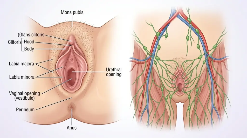
Vulvar cancer is a rare but potentially aggressive malignancy that develops on the female external genitalia—the labia majora, labia minora, clitoris, vestibule, and urethral opening. Approximately 45,240 new cases are diagnosed worldwide each year, accounting for about 4% of all gynecologic cancers. While surgical treatment alone is sufficient for most patients diagnosed at an early stage, advanced-stage cases require a multidisciplinary approach combining surgery, radiation therapy, and chemotherapy.
What Is Vulvar Cancer?
Vulvar cancer is a malignant tumor that originates in the epithelial tissue of the female external genital organs. Histologically, squamous cell carcinoma (SCC) accounts for 95% of all cases; this is followed by melanoma, basal cell carcinoma, adenocarcinoma, sarcoma, and extramammary Paget’s disease. Squamous cell carcinomas are further classified into keratinized, basaloid, and verrucous (wart-like) subtypes. The keratinized type is generally more common in postmenopausal women, while the basaloid and verrucous types are more frequently seen in younger women.
Two main biological subtypes have been identified: HPV-associated and HPV-unrelated. HPV-associated cases typically occur in women aged 35–65 with vulvar intraepithelial neoplasia (VIN) as a precursor lesion. HPV-unrelated types, on the other hand, develop in older women (ages 55–85) in association with chronic vulvar skin conditions such as lichen sclerosus. According to GLOBOCAN 2020 data, vulvar cancer accounts for 45,240 new cases and 17,427 deaths worldwide each year.
According to Assoc. Prof. Dr. Nilay Şengül, the most critical step in diagnosing vulvar cancer is for patients to notice changes in the vulvar region and not dismiss them as “signs of aging” or “temporary irritation.” While wide local excision may be sufficient for lesions detected in the early stages, more invasive surgery may be required in cases of delayed diagnosis.
Symptoms of Vulvar Cancer
The most common and persistent symptom of vulvar cancer is itching (pruritus). This is followed by vulvar bleeding, discharge, dysuria (pain during urination), and pain. However, a significant proportion of cases remain asymptomatic in the early stages, which can delay diagnosis.
The most prominent clinical finding is a mass or sore on the vulva that appears ulcerated, leukoplakic (whitish), fleshy, or wart-like. Skin discoloration (hyperpigmentation or hypopigmentation), thickening, or thinning are also early signs. Pelvic pain may occur, particularly during urination or sexual intercourse.
Assoc. Prof. Dr. Şengül emphasizes that the symptom most frequently overlooked in clinical practice is “chronic itching.” Many patients try to find temporary relief by using creams and lotions for years; however, vulvar itching that lasts longer than 6 weeks must be evaluated with a biopsy.
| Symptoms | Frequency | Severity |
|---|---|---|
| Itching (pruritus) | Most common | High — A biopsy is necessary if it lasts longer than 6 weeks |
| Vulvar bleeding | Common | High — especially in cases of postmenopausal bleeding |
| Mass/lesion | Common | Very high — biopsy required |
| Skin discoloration | Moderate | Moderate — suspected leukoplakia or erythroplakia |
| Pain / dysuria | Moderate | Moderate — may indicate an advanced stage |
What Causes Vulvar Cancer? Risk Factors
Two main pathways have been identified in the development of vulvar cancer. The first pathway is associated with HPV (human papillomavirus) infection; specifically, HPV subtypes 16 and 18 carry a risk of progression to invasive cancer following the development of vulvar intraepithelial neoplasia (VIN). Approximately 80% of untreated VIN III cases may progress to invasive vulvar cancer. This pathway is more common in young and middle-aged women (ages 35–65).
The second pathway is HPV-unrelated and is associated with chronic vulvar dermatoses (particularly lichen sclerosus), advanced age, and immunodeficiency. The severe itching caused by lichen sclerosus can lead, through the “itch-scratch cycle,” to squamous cell hyperplasia, followed by atypia and eventually cancer. This pathway is most commonly seen in women over 70 years of age.
Other important risk factors include:
- HPV infection (particularly types 16, 18, 31, and 33)
- Lichen sclerosus and other chronic vulvar dermatoses
- Advanced age (median age at diagnosis is 70)
- Smoking
- Immunodeficiency (HIV, immunosuppression following organ transplantation)
- History of cervical or vaginal cancer
- Obesity, diabetes, and hypertension (in HPV-unrelated types)
- Low socioeconomic status
According to a cohort study of 17,017 patients in Germany, the median age of patients diagnosed with vulvar cancer was 70 years (range: 19–102); 30.1% of patients were in the 70–79 age group. Patients under 55 years of age accounted for only 20.9%, while those over 75 years of age accounted for 35.8%. Younger patients are diagnosed at an earlier stage (FIGO I: 67.3%), while older patients are diagnosed at a more advanced stage (47.6%).
How Is Vulvar Cancer Diagnosed? The Diagnostic Process
The diagnosis of vulvar cancer is made through clinical examination and biopsy. Imaging methods (MRI, CT, PET-CT) do not play a role in the initial diagnosis; however, they are used in patients with advanced-stage disease to assess local spread, lymph node involvement, and distant metastases.
The diagnostic process consists of the following steps:
- Detailed medical history: The duration of itching, bleeding, discharge, pain, and previous vulvar lesions are assessed.
- Colposcopic examination: The vulvar region is examined under magnified visualization; abnormal areas are identified by applying acetic acid and Lugol’s solution.
- Biopsy: A punch biopsy is taken from the suspicious lesion; this is the gold standard for diagnosis.
- Histopathological evaluation: The tumor type, grade, depth of stromal invasion, and surgical margins are assessed.
- Imaging: Pelvic MRI (if urethral involvement is suspected), chest-abdominal CT (to check for distant metastases), and PET-CT (to assess lymph node involvement) may be ordered.
According to Assoc. Prof. Dr. Nilay Şengül, the most significant cause of diagnostic delay in vulvar cancer is patients’ reluctance to see a doctor due to feelings of shame or embarrassment. If any changes in the vulva persist for more than 2 weeks, a gynecologist should be consulted; early diagnosis directly affects treatment options and outcomes.
Stages of Vulvar Cancer and FIGO Staging
Vulvar cancer staging was revised in 2021 by the International Federation of Gynecology and Obstetrics (FIGO). This revision is based on an analysis of prospective data from 12,063 cases between 2010 and 2017 and is the first data-driven staging system in FIGO’s history.
| FIGO Stage | Definition | 5-Year Survival |
|---|---|---|
| IA | Tumor ≤2 cm, stromal invasion ≤1 mm | 86.3% |
| IB | Tumor >2 cm or stromal invasion >1 mm | 77.1% |
| II | Spread to the lower third of the urethra, vagina, or anus; negative lymph nodes | 64.5% |
| IIIA | Lymph node metastasis ≤5 mm | 60.3% |
| IIIB | Lymph node metastasis >5 mm | 49.6% |
| IIIC | Lymph node metastasis with extracapsular spread | 31.3% |
| IVA | Attachment to the pelvic bone or fixed/ulcerated lymph node | 25.9% |
| IVB | Distant metastasis | 18.3 |
According to a cohort study of 17,017 patients in Germany, the 5-year overall survival rate is 69.5% (95% CI: 68.7–70.3), with the following distribution by stage: 84.3% for FIGO I, 65.1% for FIGO II, 51.8% for FIGO III, 33.2% for FIGO IVA, and 22.2% for FIGO IVB. Lymph node status is the strongest prognostic factor: 5-year survival ranges from 70% to 93% in patients with negative nodes, while it drops to 25%–41% in patients with positive nodes.
The most significant change in the 2021 FIGO staging system is the method for measuring the depth of stromal invasion. According to the new definition, the depth of invasion is measured from the basal membrane of the deepest adjacent dysplastic rete ridge to the deepest point of invasion. In addition, the definitions of micrometastasis (>0.2 mm and ≤2 mm) and macrometastasis (>2 mm) used in cervical cancer staging have also been applied to vulvar cancer.
Vulvar Cancer Treatment
Treatment for vulvar cancer is personalized based on the stage, tumor size, histological type, lymph node status, and the patient’s overall condition. The primary treatment modality is surgery; radiation therapy and chemotherapy are used as adjuvant or neoadjuvant therapies.
Surgical Treatment
Historically, the standard treatment for vulvar cancer was radical vulvectomy combined with inguinal and pelvic lymph node dissection. However, this approach resulted in high morbidity (50% wound infection) and psychosexual problems. Today, more conservative and individualized surgical approaches are preferred.
Wide Local Excision (WLE): In localized lesions (T1, stage IA–IB), the tumor is removed with a margin of at least 1 cm of healthy tissue. Margins smaller than 8 mm have been shown to increase the risk of local recurrence. WLE is as effective as radical vulvectomy and yields better psychosexual outcomes.
Radical vulvectomy: In tumors that are larger or have spread to adjacent structures (T2, stage II), the entire vulva and tissue down to the deep fascia are removed. A modified radical vulvectomy (hemivulvectomy) may be selected.
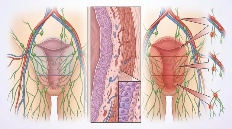
Lymph node management: Inguinofemoral lymph node dissection is the standard approach. In early-stage patients (T1b, T2) and those with clinically negative inguinal lymph nodes, sentinel lymph node (SLN) biopsy may be preferred; this reduces the risk of wound complications and lymphedema. If the SLN is positive, complete inguinofemoral lymph node dissection followed by postoperative radiation therapy is recommended.
According to Assoc. Prof. Dr. Nilay Şengül, sentinel lymph node biopsy is a revolutionary development in vulvar cancer; it significantly improves quality of life by preventing unnecessary lymph node dissection in early-stage patients. However, careful patient selection is required due to the risk of false-negative results in tumors located along the midline.
Radiotherapy
Radiotherapy is used in the following situations:
- Adjuvant radiation therapy: In the presence of 3 or more positive lymph nodes, macrometastases >10 mm, capsular rupture, or tumor at the margin (R1)
- Neoadjuvant chemoradiotherapy: To reduce tumor size prior to surgery in patients with locally advanced disease (stages III–IVA)
- Primary radiation therapy: In patients who are not candidates for surgery
The total radiation dose ranges from 60 to 70 Gy. The inguinal and pelvic regions are treated bilaterally. In recent years, the use of intensity-modulated radiation therapy (IMRT) has reduced complications by lowering the dose delivered to surrounding tissues.
Chemotherapy and Targeted Therapies
In metastatic disease (Stage IVB), systemic treatment options are limited. Platinum-based regimens (cisplatin, 5-FU, paclitaxel) are used; response rates are low. Immune checkpoint inhibitors (pembrolizumab, nivolumab) may be used in vulvar melanomas. Biological agents such as EGFR inhibitors (gefitinib, erlotinib) and trastuzumab are currently in the research phase.
Vulvar Cancer Surgery: What to Expect?
The extent of surgery for vulvar cancer varies depending on the tumor’s stage and location. While a simple wide local excision may be sufficient in early stages, radical vulvectomy and lymph node dissection may be required in advanced stages.
Preoperative: Blood tests, an EKG, a chest X-ray, and, if necessary, an anesthesia consultation are performed prior to hospital admission. Anticoagulant medications are discontinued before surgery.
Surgical Procedure: The procedure is performed under general anesthesia. The duration of surgery ranges from 2 to 5 hours, depending on the size of the tumor and the need for lymph node dissection. If a sentinel lymph node biopsy is planned, lymphatic mapping using radiocolloid and blue dye is performed before surgery.
Reconstruction: Plastic surgery reconstruction may be required to close large defects following a radical vulvectomy. Local fasciocutaneous flaps (medial thigh flap, pudendal-thigh flap) or myocutaneous flaps (rectus abdominis, gracilis) may be used. Reconstruction reduces the risk of wound dehiscence, vaginal introital stenosis, and sexual dysfunction.
Postoperative care: The hospital stay is 3–7 days. Wound care is of critical importance; the vulvar region is a moist and bacterially rich environment. The risk of wound infection is high (30–50%). Lymphedema (leg swelling) may develop in patients who undergo lymph node dissection; in such cases, physical therapy and compression stockings are recommended.
Assoc. Prof. Dr. Şengül notes that the most common problem encountered in her clinical practice following vulvar cancer surgery is “delayed wound healing.” This risk is particularly higher in patients with comorbidities such as diabetes and obesity; therefore, metabolic control should be optimized prior to surgery.
Ways to Prevent Vulvar Cancer
Prevention of vulvar cancer focuses on reducing risk factors and detecting early-stage lesions. Applicable strategies include:
- HPV vaccine: Vaccines that provide protection against HPV types 16 and 18 reduce the risk of vulvar intraepithelial neoplasia (VIN) and associated invasive cancer. While the vaccine is most effective when administered before the onset of sexual activity (ages 9–14), it can be administered up to age 45.
- Regular gynecological examinations: Performing a vulvar examination during annual gynecological checkups enables the detection of early-stage lesions. In Germany, there is an annual gynecological screening program for all women aged 20 and older, and this program plays a significant role in increasing the rate of early-stage diagnosis.
- Management of lichen sclerosus: Regular monitoring and treatment of chronic vulvar skin conditions (particularly lichen sclerosus) reduce the risk of malignant transformation.
- Smoking cessation: Smoking is an independent risk factor for both HPV-related and non-HPV-related vulvar cancers.
- Vulvar hygiene: It is recommended to avoid products containing excessive amounts of soap and perfume and to choose cotton underwear instead of synthetic options.
- Self-examination: Regular examination of the vulva using a mirror helps detect early changes.
Clinical Assessment by Assoc. Prof. Dr. Nilay Şengül
In my clinical practice, I frequently observe the following: A significant proportion of patients diagnosed with vulvar cancer experience a delay of 6–12 months between the onset of symptoms and diagnosis. The main reason for this delay is that patients interpret changes in the vulva as “signs of aging” or “temporary irritation.” According to Assoc. Prof. Dr. Nilay Şengül, vulvar itching, a lump, or a change in color lasting longer than 6 weeks must be evaluated by a gynecologist, and a biopsy should not be delayed in cases of suspicious lesions.
Furthermore, the use of sentinel lymph node biopsy has brought about a significant paradigm shift in the treatment of early-stage vulvar cancer. Today, sentinel lymph node mapping has become standard practice in patients with T1b and T2-stage disease who have clinically negative inguinal lymph nodes. This approach significantly reduces the risk of lymphedema and wound complications by preventing unnecessary lymph node dissection. However, the risk of failing to detect a sentinel lymph node is higher in tumors located along the midline and in patients with a history of previous vulvar surgery; in such cases, complete inguinofemoral lymph node dissection should be preferred.
From Diagnosis to Recovery: How Does the Process Proceed?
- Initial examination and medical history: During the initial gynecological examination, the duration of itching, bleeding, discharge, pain, and any previous vulvar lesions are thoroughly assessed. Family history, HPV vaccination status, and tobacco use are documented. The vulvar region is examined using a colposcope.
- Tests and imaging: A punch biopsy is taken from the suspicious lesion. Once a diagnosis is made, a pelvic MRI (to assess local spread), a chest-abdominal CT scan (to check for distant metastases), and, if necessary, a PET-CT scan (to assess lymph node involvement) are ordered. Blood tests (complete blood count, biochemistry, coagulation) are performed.
- Treatment planning: The stage and treatment strategy are determined at a multidisciplinary tumor board (gynecologist, medical oncologist, radiation oncologist, pathologist, radiologist). While surgery alone is sufficient in early stages, neoadjuvant chemoradiotherapy or primary surgery is planned for advanced stages.
- Surgical procedure: A wide local excision or radical vulvectomy is performed under general anesthesia. If necessary, a sentinel lymph node biopsy or inguinofemoral lymph node dissection is performed. Plastic surgery reconstruction may be performed for large defects.
- Hospital stay and discharge: The hospital stay lasts 3–7 days. Wound care, pain management, and mobilization are priorities. Patients are discharged after the drains are removed.
- Recovery and follow-up: Follow-up is recommended every 3 months for the first 3 years, every 6 months during the 4th and 5th years, and annually thereafter. Follow-up includes a vulvar examination, inguinal palpation, and imaging if necessary. Physical therapy and compression stockings are recommended for patients who develop lymphedema. Sexual function and psychological support are assessed.
According to Assoc. Prof. Dr. Nilay Şengül, the first 3 years are the most critical period for post-treatment follow-up in vulvar cancer, as data from Germany indicate that the risk of mortality is highest during this period. Regular and consistent follow-up enables the early detection of recurrence and timely intervention.
Frequently Asked Questions
Where does vulvar cancer occur?
Vulvar cancer develops on the female external genital organs, including the labia majora, labia minora, clitoris, vestibule (vaginal opening), and urethral opening. It is most commonly found in the labia majora (75%) and labia minora regions. The clitoris and perineal region are less common sites of involvement.
At what age does vulvar cancer occur?
The median age at diagnosis for vulvar cancer is 70 years (range: 19–102), and 30.1% of patients are between the ages of 70 and 79. However, HPV-related types can also occur in women aged 35–65. Patients under 55 years of age account for 20.9% of all cases, while those over 75 years of age account for 35.8%.
Does vulvar cancer cause pain?
In its early stages, vulvar cancer is generally painless; the most common symptom is itching. In advanced stages, ulceration of the tumor, spread to adjacent structures (urethra, vagina, anus), or lymph node involvement may cause pain. Dysuria (pain during urination) and pain during sexual intercourse are signs of advanced disease.
Does vulvar cancer cause back pain?
Vulvar cancer does not directly cause lower back pain. However, lower back pain may occur in advanced stages (Stage IVB) if bone metastases (to the spine or pelvic bones) develop. For this reason, bone involvement should be evaluated during imaging tests performed after diagnosis.
How long does it take for vulvar cancer to develop?
For HPV-related vulvar cancer, the time it takes for vulvar intraepithelial neoplasia (VIN) lesions to progress to invasive cancer varies; in approximately 80% of untreated VIN III cases, invasive cancer may develop over the course of several years. In HPV-unrelated types, such as chronic skin conditions like lichen sclerosus, the time to progression to cancer may be longer and less predictable; therefore, regular follow-up is important.
Is vulvar cancer contagious?
Vulvar cancer itself is not contagious. However, the underlying HPV infection in HPV-related vulvar cancers can be transmitted sexually. The HPV vaccine reduces transmission of this virus and, consequently, the risk of HPV-related vulvar cancer. Non-HPV-related types (such as those associated with lichen sclerosus) are not contagious.
Does vulvar cancer cause a foul odor in urine?
Vulvar cancer does not directly cause an odor in urine. However, tumor spread to the urethra or involvement of the urethral opening can disrupt urine flow and lead to secondary infections. A foul odor in the urine is usually associated with a urinary tract infection (UTI); a new onset of foul-smelling urine in a patient diagnosed with vulvar cancer should be evaluated for urethral involvement or a UTI.
Is vulvar cancer fatal?
The prognosis for vulvar cancer varies depending on the stage and lymph node status. In early-stage (FIGO I) disease, the 5-year survival rate is 84.3%, and this rate rises to 70–93% in lymph node-negative patients. However, in advanced-stage (FIGO IVB) disease, the 5-year survival rate drops to 18.3%. Lymph node involvement is the strongest prognostic factor; in patients with positive lymph nodes, the 5-year survival rate ranges from 25% to 41%. With early diagnosis and appropriate treatment, life expectancy can be significantly extended.
Vulvar cancer is a rare malignancy that develops in the female external genital organs but has a favorable prognosis when diagnosed early and treated. Squamous cell carcinomas account for 95% of all cases, and two main biological subtypes (HPV-associated and HPV-unrelated) have been identified. The most common symptom is itching; vulvar itching lasting longer than 6 weeks, a mass, or a change in color must be evaluated by a gynecologist. The 2021 FIGO staging system provides a more accurate prognostic classification through its data-driven approach. Surgery is the primary treatment; wide local excision is required in early stages, while a multidisciplinary approach (surgery + radiation therapy + chemotherapy) is necessary in advanced stages. Lymph node status is the factor that most strongly influences survival.
To discuss your personal situation regarding vulvar cancer and have your questions answered, you can consult with Assoc. Prof. Dr. Nilay Şengül. To schedule an appointment at her clinic at Memorial Şişli Hospital, please call 444 7 888.
References
- Alkatout I. et al., PMC/NIH, “Vulvar cancer: epidemiology, clinical presentation, and management options” — https://pmc.ncbi.nlm.nih.gov/articles/PMC4374790/
- Mack L.C. et al., BMJ Open, “Influence of stage and age on survival of patients with vulvar cancer in Germany: a retrospective study” — https://pmc.ncbi.nlm.nih.gov/articles/PMC11367380/
- Olawaiye A.B. et al., International Journal of Gynaecology & Obstetrics (FIGO), “FIGO Staging for Carcinoma of the Vulva: 2021 revision” — https://obgyn.onlinelibrary.wiley.com/doi/full/10.1002/ijgo.13880
- Singhal S. et al., World Journal of Surgical Oncology, “Patterns of care for vulvar cancer and insights from revised FIGO staging: a retrospective study” — https://link.springer.com/article/10.1186/s12957-024-03612-1
- SEER (National Cancer Institute), “Cancer Stat Facts: Vulvar Cancer” — https://seer.cancer.gov/statfacts/html/vulva.html
- ESGO (European Society of Gynecological Oncology), “Guidelines for the Management of Patients with Vulvar Cancer — Update 2023” — https://ijgc.bmj.com/content/33/6/1023