📌 What You Need to Know in a Nutshell
- Testicular cancer is the most common cancer in men aged 15–40; the 5-year survival rate is 94.6%.
- In localized disease, the 5-year survival rate reaches 99%; even in metastatic disease, it remains above 70–80%.
- BEP (bleomycin, etoposide, cisplatin) chemotherapy is the gold standard treatment for germ cell tumors.
- Undescended testicles (cryptorchidism) are the most significant risk factor; in this case, the risk increases 3- to 5-fold.
Testicular cancer is a malignancy arising from germ cells in the testicles and is the most common cancer in men aged 15–40. According to NIH/NCBI StatPearls data, it accounts for 1% of all cancers in men and 5% of urologic malignancies. According to SEER 2026 data, approximately 9,810 new cases are diagnosed annually in the United States, and the 5-year relative survival rate is 94.6%. With early diagnosis and modern treatment, it is one of the cancers with the best prognosis.
What Is Testicular Cancer?
Testicular cancer is a malignant tumor that originates from germ cells (sperm-producing cells) in the testicles. Germ cell tumors (GCTs) account for 90–95% of all testicular cancers. GCTs are divided into two main groups: seminomas and non-seminomatous germ cell tumors (NSGCTs). Seminomas account for 50–55% of GCTs and generally have a better prognosis. NSGCTs, on the other hand, include embryonal carcinoma, yolk sac tumor, choriocarcinoma, and teratoma.
Testicular cancer is one of the cancers with the highest treatment success rates. According to SEER data, the 5-year survival rate is 99% for localized disease, 96% for disease with regional lymph node involvement, and 74% even for disease with distant metastases. These high survival rates are largely due to the effectiveness of cisplatin-based chemotherapy.
According to Assoc. Prof. Dr. Nilay Şengül, the most critical step in diagnosing testicular cancer is for young men to take changes in their testicles seriously and consult a urologist without delay. According to the protocol we follow at Memorial Şişli Hospital, every patient with a suspected testicular mass undergoes an urgent scrotal ultrasound and evaluation of tumor markers (AFP, beta-hCG, LDH). This approach accelerates the diagnostic process and improves treatment success.
What Are the Symptoms of Testicular Cancer?
The symptoms of testicular cancer vary depending on the stage and histological type of the tumor. Most cases present with a painless testicular mass. Symptoms include:
- A painless, firm mass or swelling in the testicle (the most common symptom)
- A change in the size or shape of the testicle
- Scrotal pain or a feeling of heaviness
- Pain in the groin area
- Gynecomastia (breast enlargement) — in tumors that secrete beta-hCG
- Back pain (in cases of retroperitoneal lymph node involvement)
- Shortness of breath and cough (lung metastasis)
Symptoms of Testicular Cancer Metastasis
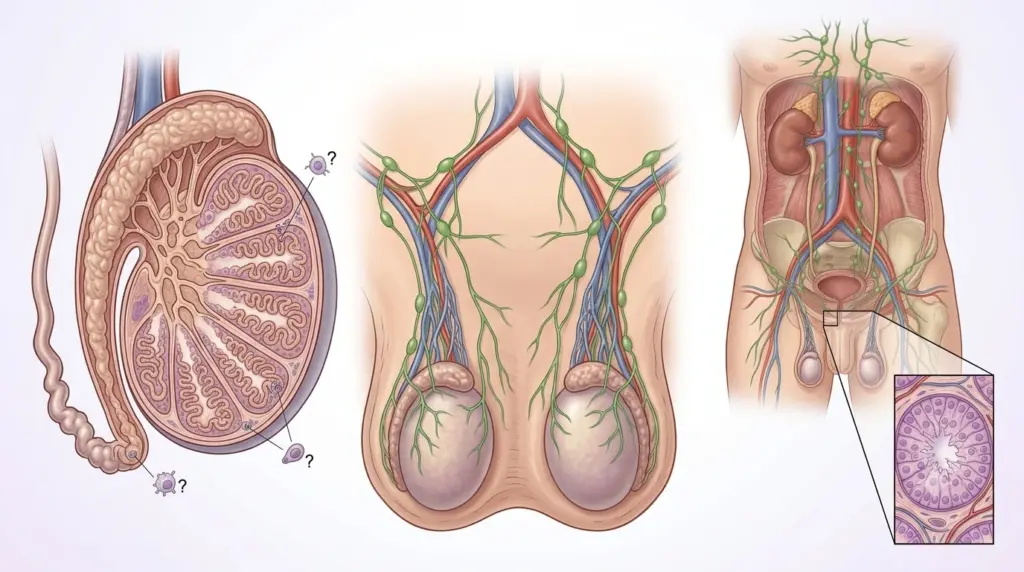
Testicular cancer metastasizes via the lymphatic system to the retroperitoneal lymph nodes and via the hematogenous route to the lungs, liver, and bones. Symptoms of metastasis vary depending on the affected organ:
Retroperitoneal lymph node involvement: Back pain (especially in the lumbar region), abdominal pain, weight loss. The retroperitoneal lymph nodes are the most common site of metastasis for testicular cancer.
Lung metastasis: Shortness of breath, cough, chest pain, hemoptysis. The lungs are the most common site of distant metastasis for testicular cancer. According to the 2024 EAU guidelines, the treatment response is generally good in patients with lung metastases.
Liver metastases: Abdominal pain, jaundice, hepatomegaly. Liver metastases are seen in advanced-stage disease.
Brain metastases: Headache, nausea, vomiting, neurological deficits. Brain metastases are rare; however, they may occur in choriocarcinoma or advanced-stage non-seminomatous germ cell tumors (NSGCT).
What Causes Testicular Cancer?
Genetic and environmental factors play a role in the development of testicular cancer. However, in most cases, the exact cause cannot be determined.
| Risk Factors | Relative Risk Increase | Explanation |
|---|---|---|
| Undescended testicle (cryptorchidism) | ~3–5 times | The most important risk factor; the risk increases in both testicles |
| Family history | ~2–3 times | History of testicular cancer in a first-degree relative |
| Previous testicular cancer | ~12–25 times | Risk of secondary cancer in the opposite testis |
| Klinefelter syndrome | ~20–50 times | Increased risk of mediastinal germ cell tumors |
| Caucasian | ~4–5 times (compared to people of African descent) | Highest incidence in the U.S. and Europe |
| Advanced age | Peak at ages 15–35 | Seminoma is more common in older men |
According to NIH/NCBI data, the incidence of testicular cancer has doubled over the past 40 years. The exact cause of this increase is unknown; however, it has been linked to environmental factors, endocrine disruptors, and lifestyle changes.
Is Testicular Cancer Fatal?
Testicular cancer is one of the cancers with the highest treatment success rates. According to SEER data, the 5-year relative survival rate is 94.6%. This rate is 99% for localized disease, 96% for regionally advanced disease, and 74% for disease with distant metastases.
According to U.S. data, the lifetime probability of a man diagnosed with testicular cancer dying from the disease is less than 1 in 5,000. Approximately 400 people die from testicular cancer each year. According to Johns Hopkins Medicine data, at any given time in the U.S., approximately 20,000 people are living with testicular cancer, and 200,000 have been cured.
The risk of death varies depending on the stage of the disease, the histological type, and the response to treatment. In patients with advanced-stage non-seminomatous germ cell tumors (NSGCT) and those with a poor prognosis, the risk of recurrence and refractory disease after treatment is higher. However, some of these patients can be cured with salvage chemotherapy and high-dose chemotherapy combined with an autologous stem cell transplant.
Stages of Testicular Cancer
Testicular cancer staging is evaluated using the AJCC TNM system in conjunction with the IGCCCG (International Germ Cell Cancer Collaborative Group) prognostic classification. The IGCCCG classification determines treatment selection and prognosis in metastatic disease.
AJCC TNM Staging
| Stage | Definition | 5-Year Survival |
|---|---|---|
| I | Tumor confined to the testis; no lymph node involvement or metastasis | 99% |
| IIA | Retroperitoneal lymph node ≤2 cm | 95–98% |
| IIB | Retroperitoneal lymph node 2–5 cm | 90–95% |
| IIC | Retroperitoneal lymph node >5 cm | 85–90% |
| III | Supradiaphragmatic lymph node or distant metastasis | 70–80% |
IGCCCG Prognostic Classification (Metastatic Disease)
The IGCCCG classification determines treatment selection and prognosis in metastatic testicular cancer. According to the 2024 EAU guidelines, this classification remains valid:
- Good prognosis: AFP <1,000 ng/mL, hCG <5,000 IU/L, LDH <1.5 times the upper limit; no non-pulmonary visceral metastases
- Intermediate prognosis: AFP 1,000–10,000 ng/mL or hCG 5,000–50,000 IU/L or LDH 1.5–10 times the upper limit; no non-pulmonary visceral metastases
- Poor prognosis: AFP >10,000 ng/mL or hCG >50,000 IU/L or LDH >10 times the upper limit; non-pulmonary visceral metastases present
According to the 2023 SEOM-GG guidelines, the standard treatment for patients with a favorable prognosis is 3 cycles of BEP or 4 cycles of EP. For patients with an intermediate or poor prognosis, 4 cycles of BEP or VIP (if bleomycin is contraindicated) are administered. Progression-free survival rates are 90% in patients with a good prognosis, 75% in those with an intermediate prognosis, and 50% in those with a poor prognosis.
Testicular Cancer Treatment
Testicular cancer treatment is personalized based on stage, histological type, and IGCCCG risk group. According to the EAU 2024 and SEOM-GG 2023 guidelines, treatment options include surgery, chemotherapy, radiation therapy, and active surveillance.
Surgical Treatment
Radical orchiectomy: This is the first step in the diagnosis and treatment of testicular cancer. Using an inguinal approach, the testis and spermatic cord are removed high up. According to the 2024 EAU guidelines, scrotal biopsy or scrotal orchiectomy is strongly discouraged, as these procedures may involve the scrotal route and inguinal lymph nodes, potentially complicating staging and treatment.
Retroperitoneal lymph node dissection (RPLND): In non-seminomatous germ cell tumors (NSGCT), this procedure is performed in patients with residual disease following chemotherapy or in marker-negative stage IIA patients. According to the 2024 EAU guidelines, nerve-sparing RPLND performed by an experienced surgeon is recommended. This procedure helps preserve ejaculatory function.
Chemotherapy
BEP (bleomycin, etoposide, cisplatin) chemotherapy is the gold standard treatment for testicular cancer. According to the 2024 EAU guidelines:
- Metastatic seminoma with a favorable prognosis: BEP x 3 or EP x 4
- Metastatic non-seminomatous germ cell tumor (NSGCT) with a favorable prognosis: BEP x 3 or EP x 4
- Metastatic disease with an intermediate or poor prognosis: BEP x 4 or VIP x 4 (if bleomycin is contraindicated)
According to the 2023 SEOM-GG guidelines, the GETUG S99 study reported a 3-year progression-free survival rate of 93% and an overall survival rate of 99% with 4 cycles of EP in seminoma with a favorable prognosis. In seminoma with an intermediate prognosis, these rates were 83% and 87%, respectively.
According to the GETUG-13 study, in poor-prognosis non-seminomatous germ cell tumors (NSGCT), individualized treatment may be applied based on the decline in tumor markers following 1 cycle of BEP. In patients with an irregular decline in tumor markers, the 5-year progression-free survival rate reached 60% with a dose-intensive protocol (compared to 47% with standard BEP).
Radiotherapy
Radiotherapy may be used as an alternative to chemotherapy in stage IIA/B seminoma. According to the 2024 EAU guidelines, for stage IIA seminoma, retroperitoneal radiation therapy or chemotherapy (BEP x 3 or EP x 4) are among the treatment options. However, chemotherapy may be preferred due to the risk of long-term cardiovascular toxicity and secondary malignancies.
Active Surveillance
In stage I seminoma and low-risk stage I NSGCT, active surveillance following radical orchiectomy is a safe option. According to the 2024 EAU guidelines, active surveillance is recommended as an alternative to adjuvant carboplatin or adjuvant radiation therapy for stage I seminoma. According to the SWENOTECA study, the 5-year recurrence rate can be reduced to less than 5% with 1 cycle of adjuvant carboplatin.
Testicular Cancer Surgery
Testicular cancer surgery includes radical orchiectomy and, when necessary, retroperitoneal lymph node dissection (RPLND). The surgical technique is determined based on the tumor stage and histological type.
Radical Orchiectomy
Using the inguinal approach, the testis and spermatic cord are removed high up. According to the 2024 EAU guidelines, the scrotal approach is strongly discouraged. The surgery takes 30–60 minutes. It can be performed under local or general anesthesia. The hospital stay is typically 1 day. A testicular prosthesis may be implanted after orchiectomy.
Retroperitoneal Lymph Node Dissection (RPLND)
In non-seminomatous germ cell tumors (NSGCT), this procedure is performed in patients with residual disease following chemotherapy or in marker-negative stage IIA patients. According to the 2024 EAU guidelines, nerve-sparing RPLND performed by an experienced surgeon is recommended. This technique preserves ejaculatory function in 90–95% of cases by protecting the sympathetic nerve sheaths while removing the retroperitoneal lymph nodes. The surgery takes 3–6 hours, and the hospital stay is 3–7 days.
Assoc. Prof. Dr. Şengül emphasizes that in her clinical practice, temporary ejaculatory dysfunction may develop in 10–15% of patients following RPLND; however, this rate can be reduced to below 5% with a nerve-sparing technique. For this reason, she stresses that it is critical for RPLND to be performed at an experienced center.
Clinical Assessment by Assoc. Prof. Dr. Nilay Şengül
In my clinical practice, I frequently observe the following: Young male patients diagnosed with testicular cancer often consider the changes in their testicles to be “normal” prior to diagnosis and do not seek medical attention for an average of 2–3 months. In the cases we evaluate at Memorial Şişli Hospital, we see that this delay leads to disease progression and a more complicated treatment plan.
According to Assoc. Prof. Dr. Nilay Şengül, the most significant paradigm shift in testicular cancer has been the transition from the “same dose of chemotherapy for every metastatic patient” approach to “individualized treatment based on the IGCCCG risk group and tumor markers.” In particular, the GETUG-13 study has demonstrated that intensifying treatment based on a decrease in tumor markers after one cycle of BEP may be beneficial for patients with a poor prognosis. This approach enhances treatment efficacy while minimizing unnecessary toxicity. Assoc. Prof. Dr. Şengül also emphasizes that, in stage I seminoma, an active surveillance strategy is equivalent to adjuvant therapy in terms of long-term survival; however, it must be selected based on patient-specific risk factors.
From Diagnosis to Recovery: How Does the Process Proceed?
- Initial Examination and Evaluation: The patient’s symptoms (testicular mass, pain, swelling), family history (cryptorchidism, history of testicular cancer), and general health status are assessed. During the physical examination, the testicles are palpated, and the lymph node regions and gynecomastia are assessed. Assoc. Prof. Dr. Şengül emphasizes that, in her clinical practice, it is critical for young men to develop the habit of performing testicular self-examination (TSE) for early diagnosis.
- Diagnostic Tests:
- Scrotal ultrasound: Determines whether a testicular mass is solid or cystic, its size, and whether it involves the epididymis; mandatory for diagnosis
- Tumor markers: AFP (alpha-fetoprotein), beta-hCG (beta human chorionic gonadotropin), LDH (lactate dehydrogenase); used for staging and monitoring treatment response
- CT (chest + abdomen + pelvis): Assesses lymph node involvement and lung metastases; mandatory for staging
- PET-CT: Used to assess the presence of viable tumor in residual seminoma masses (≥3 cm)
- Radical Orchiectomy: The testis and spermatic cord are removed via an inguinal approach. Preoperative sperm cryopreservation is recommended. The pathology report specifies the tumor type, size, venous/lymphatic invasion, and rete testis involvement.
- Staging and Treatment Planning: AJCC TNM staging and IGCCCG risk classification are applied. An individualized treatment plan is developed during a multidisciplinary tumor board meeting (urology, medical oncology, radiation oncology, pathology). According to the 2024 EAU guidelines, options such as active surveillance, adjuvant carboplatin, or radiation therapy are discussed for stage I seminoma.
- Treatment Implementation:
- Stage I: Active surveillance or adjuvant therapy (seminoma: carboplatin or radiation therapy; NSGCT: active surveillance or BEP × 1)
- Stages II–III (good prognosis): BEP × 3 or EP × 4
- Stage II–III (intermediate/poor prognosis): BEP × 4 or VIP × 4
- Residual mass after chemotherapy: RPLND (NSGCT) or evaluation with PET-CT (seminoma)
- Follow-up and Monitoring: Checkups are scheduled every 3–6 months for the first 2 years, every 6–12 months for the next 3 years, and annually starting in the 5th year. Each checkup includes a physical examination, tumor marker testing, and imaging. Long-term follow-up should include cardiovascular evaluation, pulmonary function tests, and screening for secondary malignancies.
Frequently Asked Questions
How does testicular cancer develop?
Testicular cancer develops as a result of genetic mutations in the germ cells of the testicles. Cryptorchidism (undescended testicle), family history, a previous history of testicular cancer, and Klinefelter syndrome increase the risk. In most cases, the exact cause cannot be determined. According to NIH/NCBI data, the incidence has doubled over the past 40 years; however, the reason for this increase is not fully understood.
How does testicular cancer develop?
Testicular cancer develops as a result of uncontrolled cell growth caused by chromosomal abnormalities in germ cells (particularly 12p chromosome amplification). This process may begin during fetal development and become clinically apparent years later. An undescended testis leads to abnormal development of germ cells, increasing the risk by 3 to 5 times. Environmental factors (endocrine disruptors, pesticides) and genetic predisposition may also play a role.
How is testicular cancer diagnosed?
A definitive diagnosis is made through pathological examination following a radical orchiectomy. However, clinically, a scrotal ultrasound determines whether the testicular mass is solid or cystic. Elevated tumor markers (AFP, beta-hCG, LDH) support the diagnosis of malignancy. Staging is performed using CT. Regular self-examination of the testicles (TSE) by young men is critical for early diagnosis. If there is a painless testicular mass, scrotal pain lasting longer than 4 weeks, or a change in testicular size, a urologist should be consulted immediately.
Is testicular cancer contagious?
No, testicular cancer is not contagious. Cancer cells do not spread from one person to another. However, certain infections (such as HPV) can increase the risk of cancer; these infections can be transmitted sexually. Testicular cancer itself is not contagious and does not run in families; however, a genetic predisposition (family history) can increase the risk.
Where does testicular cancer occur?
Testicular cancer originates in the germ cells of the testicles. It is most commonly found in the right testicle; however, it can occur equally in both testicles. It spreads via the lymphatic system to the retroperitoneal lymph nodes (paraaortic, paracaval). It metastasizes via the hematogenous route to the lungs, liver, bones, and, rarely, the brain. Extragonadal germ cell tumors may also be found in the mediastinum and retroperitoneum.
Where does testicular cancer spread?
Testicular cancer spreads primarily through the lymphatic system. The first stop is the retroperitoneal lymph nodes (paraaortic, paracaval). Tumors in the right testis spread to the lymph nodes in front of the inferior vena cava, while tumors in the left testis spread to the lymph nodes in front of the aorta. Subsequently, the supradiaphragmatic lymph nodes and lungs are involved. Hematogenous metastases can occur in the lungs, liver, bones, and brain. Choriocarcinoma exhibits early and aggressive hematogenous spread.
Can testicular cancer be detected on an ultrasound?
Yes, scrotal ultrasound is the first and most important imaging method for diagnosing testicular cancer. Ultrasound determines whether the mass is solid or cystic, its size, the extent of extratesticular involvement, and the condition of the contralateral testis. Solid, hypoechoic masses suggest malignancy. Microlithiasis (testicular microliths) may increase the risk of testicular cancer; however, it is not a sign of malignancy on its own. Ultrasound provides 90–95% accuracy in establishing a diagnosis prior to biopsy.
Can testicular cancer be detected through blood tests?
Serum tumor markers (AFP, beta-hCG, LDH) are used in the diagnosis and monitoring of testicular cancer. AFP and beta-hCG may be elevated in non-seminomatous germ cell tumors (NSGCT); in seminoma, beta-hCG may be mildly elevated, while AFP remains normal. LDH indicates tumor burden and cell turnover rate. These markers play a critical role in staging, assessing treatment response, and monitoring for recurrence. However, cancer may still be present even if the markers are normal; therefore, imaging and pathology are mandatory.
How quickly does testicular cancer spread?
The rate at which testicular cancer spreads varies depending on the histological subtype. Seminomas generally grow more slowly and spread via the lymphatic system. NSGCTs (particularly choriocarcinoma and embryonal carcinoma) may behave more aggressively and can spread hematogenously. In localized Stage I disease, lymph node involvement may develop within months. However, testicular cancer can be controlled with early diagnosis and treatment; therefore, it is important to seek medical attention without delay if suspicious symptoms arise.
How many years does it take for testicular cancer to be fatal?
When treated appropriately, the risk of death from testicular cancer is low. According to SEER data, the 5-year relative survival rate is 94.6%. It is 99% for localized disease and 74% even for metastatic disease. The prognosis worsens in untreated or treatment-resistant cases. In the U.S., the probability of a man diagnosed with testicular cancer dying from the disease is less than 1 in 5,000. Approximately 400 people die from testicular cancer each year. Early diagnosis and BEP chemotherapy significantly improve survival rates.
What helps with testicular cancer?
Testicular cancer treatment is personalized based on stage and risk group. Radical orchiectomy is the first step in diagnosis and treatment. BEP chemotherapy is the gold standard for metastatic disease. Radiation therapy may be used as an alternative for stage IIA/B seminoma. Active surveillance is a safe option for low-risk stage I patients. Regular follow-up after treatment is important for monitoring cardiovascular health, lung function, and screening for secondary cancers. According to Assoc. Prof. Dr. Şengül, patients receiving psychosocial support after treatment is also critical for their quality of life.
Does testicular cancer recur?
Yes, it can recur. In stage I seminoma, the 5-year recurrence rate is 15–20% with active surveillance and less than 5% with adjuvant carboplatin. In low-risk stage I NSGCT patients, the recurrence rate is 15–30%. In metastatic disease, the complete response rate after BEP chemotherapy is 80–90%; however, recurrence or refractory disease may develop in 10–20% of cases. Recurrence is most common within the first 2 years. Salvage chemotherapy and high-dose chemotherapy plus autologous stem cell transplant offer a chance of cure in cases of recurrence or refractory disease.
Which department treats testicular cancer?
A multidisciplinary approach is used in the diagnosis and treatment of testicular cancer. The physician who makes the diagnosis and performs the initial evaluation is typically a urologist. Medical oncology, radiation oncology, and urology teams are involved in treatment planning. According to the 2024 EAU guidelines, all testicular cancer patients should be evaluated at an experienced center. According to 2024 ESMO data, high-risk and recurrent/refractory patients should be referred to experienced centers.
Testicular cancer is the most common cancer among men aged 15–40 and is one of the malignancies with the highest survival rates when diagnosed early and treated with modern therapies. Five-year survival reaches 99% in localized disease; even in metastatic disease, it exceeds 70–80%. BEP chemotherapy is the gold standard treatment for germ cell tumors. Radical orchiectomy is the first step in diagnosis and treatment. It is critically important for young men to develop the habit of performing self-examinations of their testicles to ensure early diagnosis and successful treatment.
To discuss your personal situation regarding testicular cancer and have your questions answered, you can consult with Assoc. Prof. Dr. Nilay Şengül. To schedule an appointment at her clinic at Memorial Şişli Hospital, please call 444 7 888.
Sources
- NIH/NCBI StatPearls, "Testicular Cancer" — https://www.ncbi.nlm.nih.gov/books/NBK563159/
- SEER Cancer Stat Facts: Testicular Cancer — https://seer.cancer.gov/statfacts/html/testis.html
- EAU Guidelines on Testicular Cancer 2024 — https://d56bochluxqnz.cloudfront.net/documents/EAU-Guidelines-on-Testicular-Cancer-2024.pdf
- SEOM-GG Clinical Guidelines for the Management of Germ-Cell Testicular Cancer (2023) — https://pmc.ncbi.nlm.nih.gov/articles/PMC11467073/
- Johns Hopkins Medicine, "Testicular Cancer Statistics" — https://www.hopkinsmedicine.org/health/conditions-and-diseases/testicular-cancer/testicular-cancer-statistics
- ESMO 2024: Controversies in Testicular Cancer: Escalation or De-escalation? — https://www.urotoday.com/conference-highlights/esmo-2024/esmo-2024-testicular-and-penile-cancer/154951-esmo-2024-controversies-in-testis-cancer-escalation-or-de-escalation.html
- CA: A Cancer Journal for Clinicians, "Cancer Statistics, 2025" — https://doi.org/10.3322/caac.21871