📌 What You Need to Know in a Nutshell
- Soft tissue cancer (sarcoma) accounts for 1% of cancers in adults; there are more than 50 different subtypes.
- In localized stages, 5-year survival ranges from 60% to 80% with surgical resection; in advanced stages, median survival is 12–18 months.
- According to the SU2C-SARC032 study published in The Lancet in 2024, the combination of pembrolizumab, radiation therapy, and surgery increased 2-year disease-free survival from 52% to 67%.
- Histological grade and tumor size are the most important prognostic factors; lymph node involvement is rare (5%).
Soft tissue cancer (sarcoma) is a rare group of malignant tumors originating from connective tissue, muscle, fat, blood vessels, nerves, and subcutaneous tissue outside the skeleton. It accounts for approximately 1% of cancers in adults and has more than 50 different histological subtypes. Early diagnosis and appropriate treatment reduce both the risk of local recurrence and the risk of distant metastasis.
What Is Soft Tissue Cancer (Sarcoma)?
Soft tissue cancer (sarcoma) is a group of malignant neoplasms that originate from mesenchymal cells and can affect all supporting tissues other than bone. The most common subtypes are liposarcoma (adipose tissue), leiomyosarcoma (smooth muscle), synovial sarcoma, angiosarcoma (vascular tissue), and pleomorphic sarcoma of undetermined differentiation (UPS). Unlike carcinomas (cancers of epithelial origin), sarcomas are rarer, and their biological behavior varies depending on the subtype.
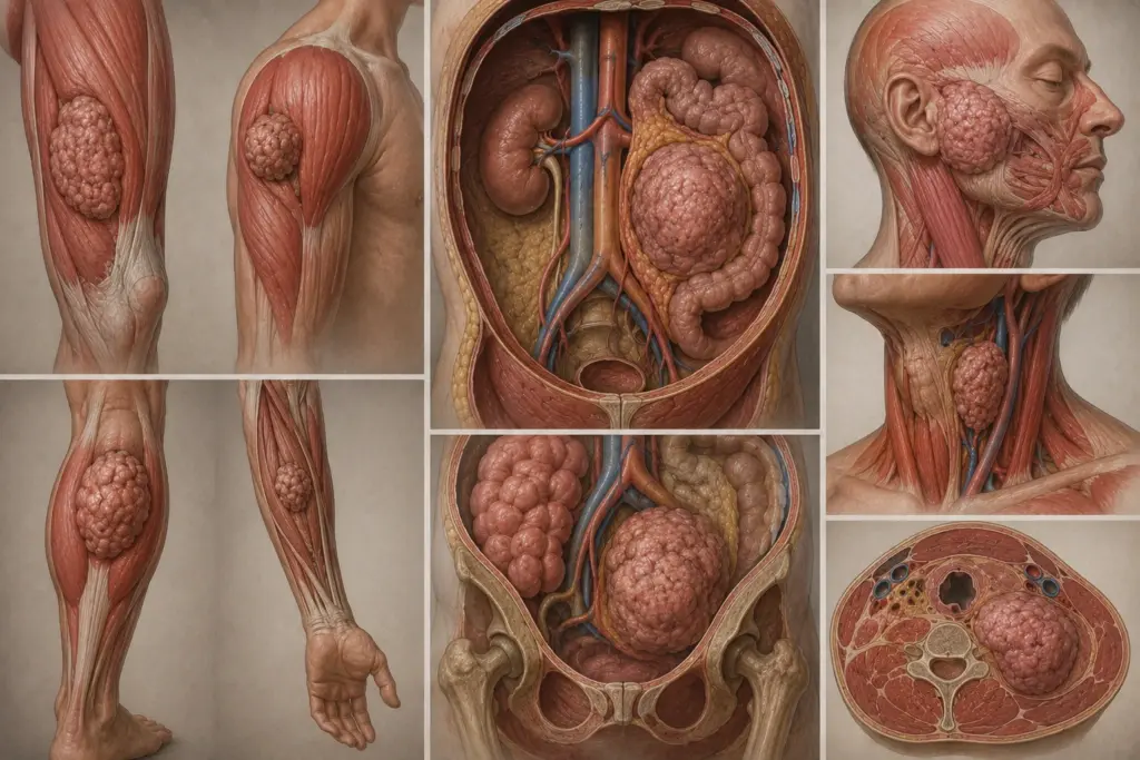
According to NIH/NCBI StatPearls data, sarcomas account for 1% of adult cancers; however, they account for 15–20% of childhood cancers. The median age at diagnosis is between 50 and 60 years. They are slightly more common in men than in women. They are most frequently found in the extremities (43%), the retroperitoneal region (15%), the head and neck region (9%), and the trunk (30%).
According to Assoc. Prof. Dr. Nilay Şengül, the most critical factor in the diagnosis and treatment of sarcomas is ensuring that the patient undergoes a multidisciplinary evaluation at an experienced sarcoma center. According to the protocol we follow at Memorial Şişli Hospital, detailed imaging prior to biopsy and determination of the subtype via immunohistochemistry after biopsy are mandatory for every patient with suspected sarcoma. An incorrect diagnosis or inadequate surgery significantly increases the risk of local recurrence and distant metastasis.
What Causes Soft Tissue Cancer?
The exact cause of sarcomas is unknown in most cases. However, associations with certain risk factors and genetic syndromes have been identified.
Major Risk Factors
Radiation: The development of sarcomas in areas previously treated with radiation therapy is one of the best-known risk factors. Radiation-induced sarcomas typically emerge 10–15 years after the initial treatment.
Chemical Exposure: A link has been reported between chemicals such as herbicides, chlorophenols, and vinyl chloride and angiosarcoma. Lymphedema (particularly following breast cancer) increases the risk of angiosarcoma.
Genetic syndromes: Neurofibromatosis type 1 (NF1) increases the risk of malignant peripheral nerve sheath tumors (MPNSTs). Li-Fraumeni syndrome (TP53 mutation) is associated with childhood sarcomas. Retinoblastoma, Gardner syndrome, and familial adenomatous polyposis are other genetic conditions that increase the risk of sarcoma.
Lymphedema: Chronic lymphedema (Stewart-Treves syndrome) significantly increases the risk of angiosarcoma. This risk is particularly observed in patients with lymphedema following mastectomy.
| Risk Factor | Associated Sarcoma Subtype | Relative Risk Increase |
|---|---|---|
| Previous Radiotherapy | Radiation-induced sarcoma | ~0.1–0.5% (10–15 years later) |
| Neurofibromatosis type 1 | Malignant peripheral nerve sheath tumor | ~10–15% lifetime risk |
| Chronic lymphedema | Angiosarcoma | High (Stewart-Treves) |
| Chemical exposure | Angiosarcoma, hepatic sarcoma | Occupational exposure-related |
Soft Tissue Cancer Symptoms
Symptoms of sarcoma vary depending on the tumor’s location, size, and subtype. Sarcomas of the extremities are the most common type.
- A painless, slowly growing subcutaneous mass (the most common initial symptom)
- Redness, increased warmth, or venous dilation of the skin over the mass
- Pain (resulting from nerve compression or muscle invasion)
- Limited range of motion (if located near a joint)
- Weight loss, fatigue, and night sweats (advanced stage)
- Shortness of breath and cough (lung metastasis)
Symptoms of Soft Tissue Cancer in the Leg
The leg is the most common site for sarcomas (43%). The back of the thigh (popliteal fossa) and the inner thigh are the most common sites of occurrence. In leg sarcomas, the mass usually begins painlessly; however, as it grows, it can cause difficulty walking and limit movement in the knee or hip. Venous thrombosis or lymphedema may develop. Unlike retroperitoneal sarcomas, leg sarcomas are detected earlier because the mass is palpable and visible.
Symptoms of Soft Tissue Cancer in the Arm
Arm sarcomas are less common than leg sarcomas. The shoulder, upper arm, and elbow area are the most common sites. In arm sarcomas, the mass may restrict shoulder or elbow movement. Numbness or weakness in the hand and fingers may develop as a result of nerve compression. Neurological symptoms tend to appear earlier, particularly in synovial sarcoma and MPNST, as these tumors originate from nerve sheaths.
Stages of Soft Tissue Cancer
Sarcoma staging is performed according to the AJCC 8th edition TNM system. In this system, tumor size (T), lymph node involvement (N), distant metastasis (M), and histological grade (G) are evaluated. NCCN guidelines follow the AJCC/UICC staging system; ESMO, on the other hand, recommends the use of validated nomograms.
| Stage | TNM Definition | 5-Year Survival |
|---|---|---|
| IA | T1 (<5 cm), N0, M0, G1 | 80–90% |
| IB | T2–T4 (>5 cm), N0, M0, G1 | 70–80% |
| II | T1, N0, M0, G2–G3 | 60–70 |
| IIIA | T2, N0, M0, G2–G3 | 50–60 |
| IIIB | T3–T4, N0, M0, G2–G3 | 40–50% |
| IV | Any T, N1 or M1, Any G | 10–20% |
According to NIH/NCBI data, histological grade and tumor size are the most important prognostic factors. Histological grade predicts distant metastasis and survival, while tumor size predicts local recurrence and distant metastasis. The Memorial Sloan Kettering sarcoma nomogram predicts the 3- and 5-year risk of local recurrence using grade, depth, size, margin status, age, and histology.
Soft Tissue Cancer Treatment
Sarcoma treatment is personalized based on the tumor’s stage, subtype, location, and the patient’s performance status. According to NCCN and ESMO guidelines, a multidisciplinary evaluation at an experienced sarcoma center is essential for treatment planning.
Surgical Treatment
Surgical resection is the primary treatment for localized sarcomas. According to NCCN guidelines, a multidisciplinary team should evaluate the case before amputation is considered. The goal is to achieve a surgical margin of >1 cm or an intact fascial plane. According to ESMO guidelines, the standard surgical procedure is en bloc excision with an R0 margin; however, R1 excision is acceptable in selected cases.
In extremity sarcomas, limb-sparing surgery is the gold standard. Radical compartment resection is not routinely necessary and does not provide a survival benefit. Narrow margins may be required to preserve critical structures; in such cases, function and quality of life are prioritized. According to NCCN guidelines, evaluation must be performed at a center with experience in sarcoma prior to amputation.
Radiation Therapy
Radiation therapy is administered as adjuvant or neoadjuvant therapy. According to NCCN guidelines, preoperative and postoperative radiation therapy is a Category 1 recommendation for patients with Stage IIB–III disease. According to data from randomized trials, radiation therapy improves disease-free survival after surgery (but does not affect overall survival).
Preoperative radiation therapy carries a lower risk of late toxicity (fibrosis, edema, joint stiffness) compared to postoperative radiation therapy. For this reason, the NCCN panel prefers preoperative radiation therapy, particularly for large treatment volumes. The radiation therapy dose and fractionation are determined by an experienced radiation oncologist based on normal tissue constraints.
Chemotherapy
Anthracycline-based chemotherapy (doxorubicin) is used as first-line treatment for advanced-stage sarcomas. According to ESMO guidelines, the combinations of doxorubicin + ifosfamide or doxorubicin + dacarbazine are preferred when the potential for response is favorable and the patient’s performance status is good. Response rates range from 10% to 50%; these rates vary depending on the histological subtype, grade, and the individual patient.
Trabectedin is a Category 1 recommendation for second-line and subsequent treatment in liposarcoma and leiomyosarcoma. Pazopanib is a second-line option for non-adipogenic sarcomas. Eribulin is a Category 1 option for liposarcomas and a Category 2A option for other subtypes.
Immunotherapy and Targeted Therapies
Immune checkpoint inhibitors (ICIs) have emerged as a promising area in sarcoma treatment. In the SARC028 Phase II trial, the objective response rate with pembrolizumab monotherapy was found to be 18% in sarcomas. The best responses were observed in pleomorphic sarcoma of undetermined differentiation (23%) and dedifferentiated liposarcoma (10%).
The SU2C-SARC032 study, published in *The Lancet* in 2024, represents a significant turning point in sarcoma treatment. In this randomized Phase III trial, among patients with Stage III undifferentiated pleomorphic sarcoma and pleomorphic/dedifferentiated liposarcoma, the addition of pembrolizumab to preoperative radiation therapy plus surgery increased 2-year disease-free survival from 52% to 67% (HR: 0.61; 90% CI: 0.39–0.96). This benefit is more pronounced in patients with Grade 3 sarcoma (HR: 0.47; 95% CI: 0.25–0.89).
Larotrectinib and entrectinib are the standard treatments for NTRK-fusion-positive sarcomas. Imatinib is the standard medical treatment for dermatofibrosarcoma protuberans (DFSP). The combination of pembrolizumab and lenvatinib has been reported to provide a median progression-free survival of 7.8 months in undifferentiated pleomorphic sarcoma.
[VISUAL SUGGESTION: Sarcoma treatment algorithm diagram — showing surgical, radiation therapy, chemotherapy, and immunotherapy options by stage and subtype — Alt text: "Stage-based treatment algorithm decision tree diagram for soft tissue cancer"]
Soft Tissue Cancer Surgery
Sarcoma surgery aims to remove the tumor with clear margins of healthy tissue and preserve limb function. The surgical technique is determined based on the tumor’s size, location, and subtype.
Surgical Techniques
Limb-sparing surgery: The tumor is removed along with surrounding healthy tissue (margin). The margin width varies depending on the subtype; for example, a margin of 1 cm or less is sufficient for atypical lipomatous tumors or well-differentiated liposarcomas, whereas much wider resection is required for locally invasive subtypes such as dermatofibrosarcoma protuberans, myxofibrosarcoma, or angiosarcoma.
Retroperitoneal sarcoma surgery: R0 resection is the goal; however, an R1 margin is acceptable to preserve critical structures. According to NCCN guidelines, surgery with or without intraoperative radiation therapy may be performed for resectable disease. A radical compartment approach is not routinely required.
Amputation: This is the last resort when limb-sparing surgery is not possible. According to NCCN guidelines, evaluation at a center with experience in sarcoma is mandatory prior to amputation.
Soft Tissue Cancer Mortality Rate
The mortality rate for sarcomas varies depending on stage, grade, subtype, and response to treatment. According to NIH/NCBI data, curative resection can provide good long-term survival in localized and early-stage lesions; however, recurrences are common. Most recurrences occur within the first 5 years; however, the risk persists even 10–15 years later.
In advanced-stage sarcomas, a cure is not possible, and median survival is 12–18 months. According to a 2024 study in *Frontiers in Endocrinology*, the results of multivariate analysis in patients with metastatic sarcoma are as follows:
- Age >65: HR 1.28 (95% CI: 1.10–1.49)
- Pathological grade III–IV: HR 1.87 (95% CI: 1.43–2.45)
- Lung metastasis: HR 1.82 (95% CI: 0.52–6.35)
- Brain metastases: HR 2.03 (95% CI: 0.61–6.74)
- Bone metastasis: HR 1.90 (95% CI: 0.54–6.66)
- Underwent surgery: HR 0.55 (95% CI: 0.47–0.64)
- Received chemotherapy: HR 0.58 (95% CI: 0.50–0.67)
The chemotherapy response rate in sarcomas ranges from 10% to 50%; this rate varies depending on the histological subtype, grade, and the individual patient. Immunotherapy and targeted therapies have improved survival in certain subtypes in recent years.
Clinical Assessment by Assoc. Prof. Dr. Nilay Şengül
In my clinical practice, I frequently observe the following: Patients diagnosed with sarcoma typically spend an average of 3–6 months consulting various medical specialties prior to diagnosis, and this process leads to a delay in the initiation of treatment. With the multidisciplinary sarcoma protocol we implement at Memorial Şişli Hospital, we aim to reduce this period to 2–4 weeks.
According to Assoc. Prof. Dr. Nilay Şengül, the most significant paradigm shift in sarcoma treatment is the transition from a “one-size-fits-all” approach to “histology-specific personalized treatment.” In particular, the SU2C-SARC032 study published in *The Lancet* in 2024 demonstrated that pembrolizumab significantly extended progression-free survival in patients with stage III undifferentiated pleomorphic sarcoma and pleomorphic liposarcoma. This finding reinforces the role of immunotherapy in sarcoma treatment. Assoc. Prof. Dr. Şengül emphasizes that accurately determining the sarcoma subtype in clinical practice directly influences treatment selection and prognosis.
From Diagnosis to Recovery: How Does the Process Proceed?
- Initial Examination and Evaluation: The patient’s symptoms (mass, pain, limited mobility), family history, and general health status are assessed. During the physical examination, the mass’s size, mobility, skin changes, and lymph nodes are assessed. Assoc. Prof. Dr. Şengül particularly emphasizes asking about the mass’s growth rate and pain characteristics during the initial consultation in her clinical practice.
- Diagnostic Tests:
- MRI (magnetic resonance imaging): This is the primary imaging modality for extremity sarcomas; it assesses tumor size, depth, and its relationship to muscles and nerves.
- CT (computed tomography): Used to screen for lung metastases; serves as an alternative to MRI for calcified lesions and retroperitoneal sarcomas
- PET-CT: Useful for evaluating the response to neoadjuvant chemotherapy in large (>3 cm) and deep-seated tumors
- Biopsy: Core needle biopsy (preferred) or open incisional biopsy; the biopsy site should be positioned parallel to the axis of the future resection
- Staging and Risk Assessment: The AJCC 8th edition TNM staging system is applied. Histological grade (FNCLCC system), tumor size, depth, and margin status are entered into prognostic nomograms. The Memorial Sloan Kettering nomogram predicts the 3- and 5-year risk of local recurrence.
- Treatment Approach:
- Stage IA–IB (low grade): Surgical resection; if the margin is ≤1 cm and the fascia is not intact, re-resection or radiation therapy
- Stage II–III (high-grade): Surgery plus preoperative or postoperative radiation therapy; adjuvant chemotherapy may be considered for stages IIB and III
- Stage IV (metastatic): Systemic therapy (chemotherapy, immunotherapy, targeted therapy); metastasectomy in patients with oligometastases
- Hospital Stay and Discharge: For limb-sparing surgery, the hospital stay is 3–7 days; for amputation, it is 5–10 days. In retroperitoneal surgery, this period may extend to 7–14 days. Rehabilitation and physical therapy are planned following discharge.
- Follow-up and Monitoring: According to NCCN guidelines, physical examinations are performed every 3–6 months for the first 2–3 years, followed by annual follow-up. Chest imaging and MRI/CT scans of the primary site are scheduled based on histology. The likelihood of recurrence is low after 10 years; however, follow-up should be tailored to the specific subtype.
Frequently Asked Questions
What is the life expectancy for soft tissue cancer?
Life expectancy varies depending on stage, grade, and subtype. In localized stages (Stages I–III), 5-year survival ranges from 60% to 80% with surgery plus radiation therapy. In advanced (metastatic) stages, median survival is 12–18 months. According to a 2024 Lancet study, in Stage III pleomorphic sarcoma of undetermined differentiation, 2-year disease-free survival reached 67% with pembrolizumab plus radiation therapy plus surgery. Histological grade is the most important prognostic factor.
Can soft tissue cancer be fatal?
In sarcomas diagnosed at an early stage (Stages I–II) and treated appropriately, the cure rate is high. However, the prognosis worsens in high-grade, large-size, or metastatic sarcomas. The risk of recurrence persists even 10–15 years later; therefore, long-term follow-up is crucial. A cure is not possible in metastatic disease; however, systemic treatments can prolong survival.
What is the life expectancy for soft tissue cancer?
In localized sarcomas, the 5-year survival rate is 60–80%, while in metastatic sarcomas, the median survival is 12–18 months. According to the Frontiers 2024 study, the survival risk for patients with metastatic sarcoma who underwent surgery is 45% lower than for those who did not (HR: 0.55). Patients receiving chemotherapy also have a similar survival advantage (HR: 0.58). Age over 65, lung/brain/bone metastases, and high-grade tumors are prognostic factors.
Which department treats soft tissue cancer?
A multidisciplinary approach is used in the diagnosis and treatment of sarcoma. The physician who makes the diagnosis and performs the initial evaluation is typically a specialist in orthopedic oncology or general surgery. Treatment planning involves teams from medical oncology, radiation oncology, orthopedic oncology, and pathology. According to ESMO guidelines, all sarcoma patients should be referred to a sarcoma center or network.
How is soft tissue cancer diagnosed?
A definitive diagnosis is made via biopsy. However, during the clinical examination, the physician assesses the mass’s size, mobility, skin changes, and lymph nodes. An MRI shows the tumor’s relationship to muscle, nerves, and bone. CT is used to screen for lung metastases. PET-CT is useful for evaluating the response to neoadjuvant chemotherapy. A biopsy is performed via core needle biopsy (preferred) or open incisional biopsy; the biopsy site should be positioned parallel to the future surgical axis.
What are the early symptoms of soft tissue cancer?
The most common initial symptom is a painless, slowly growing subcutaneous mass. The mass may grow over time and cause pain. Skin redness, increased warmth, or venous dilation may be observed. If the tumor is located near a joint, limited mobility may develop. In retroperitoneal sarcomas, lower back pain, weight loss, and an abdominal mass are more common symptoms. Any subcutaneous mass that grows within 4–6 weeks must be evaluated.
How long do patients with soft tissue cancer live?
The answer to this question depends on the stage of the disease and the response to treatment. In localized, low-grade sarcomas, 10-year survival can reach 70–80%. In high-grade, locally advanced sarcomas, 5-year survival is 40–50%. In metastatic disease, median survival is 12–18 months. Survival is significantly prolonged in metastatic patients who undergo surgery and chemotherapy (HR: 0.55 and 0.58). Immunotherapy improves survival in certain subtypes (UPS, liposarcoma).
Is there a treatment for soft tissue cancer?
Yes, there is a treatment. Surgical resection is the primary treatment for localized disease; radiation therapy and chemotherapy are used as adjuvant therapies. According to a 2024 Lancet study, the combination of pembrolizumab, radiation therapy, and surgery in stage III UPS and liposarcoma increased 2-year disease-free survival to 67%. For metastatic disease, doxorubicin-based chemotherapy, immunotherapy, and targeted therapies (pazopanib, trabectedin, eribulin) are used. Larotrectinib is the standard treatment for patients with NTRK fusion-positive tumors.
Where does soft tissue cancer occur?
It is most commonly found in the extremities (43%), particularly the thigh and popliteal fossa. The retroperitoneal space (15%), the body wall (30%), and the head and neck (9%) are other common sites. The location varies by subtype: liposarcoma is more common in the retroperitoneal space; synovial sarcoma in the joints of the extremities; angiosarcoma in the skin and soft tissues; and rhabdomyosarcoma is more common in the head and neck and the genitourinary system in children.
Is soft tissue cancer genetic?
Most sarcomas develop sporadically (randomly); however, certain genetic syndromes increase the risk. Neurofibromatosis type 1 (NF1) increases the risk of malignant peripheral nerve sheath tumors (MPNSTs). Li-Fraumeni syndrome (TP53 mutation) is associated with childhood sarcomas. Retinoblastoma (RB1 mutation), Gardner syndrome, and familial adenomatous polyposis also lead to an increased risk. Genetic counseling is recommended for patients with a family history.
How is soft tissue cancer diagnosed?
A definitive diagnosis is made by biopsy. MRI shows the tumor’s size and its relationship to surrounding tissues. CT is used to screen for lung metastases. Immunohistochemistry (IHC) and conventional cytogenetic analyses of biopsy specimens play a critical role in determining the subtype. NTRK fusions, PD-L1 expression, and tumor mutation burden (TMB) aid in the selection of targeted therapy and immunotherapy. PET-CT is used to evaluate the response to neoadjuvant therapy.
Who is affected by soft tissue cancer?
Sarcomas can occur at any age; however, the median age at diagnosis is 50–60 years. Some subtypes vary by age: rhabdomyosarcoma is more common in childhood, synovial sarcoma in young adults (ages 15–35), and pleomorphic liposarcoma and UPS are more common in middle-aged and older adults. Men are slightly more likely to be affected than women. A history of radiation exposure, genetic syndromes, and chronic lymphedema are risk factors.
Soft tissue cancer (sarcoma) is a rare but heterogeneous group of cancers. Early-stage diagnosis and treatment at an experienced sarcoma center reduce both the risk of local recurrence and the risk of distant metastasis. Surgical resection is the gold standard for localized disease; radiation therapy and chemotherapy are used as adjuvant treatments. The SU2C-SARC032 study, published in *The Lancet* in 2024, demonstrated that pembrolizumab significantly extended progression-free survival in stage III UPS and liposarcoma, ushering in a new era in sarcoma treatment.
To discuss your personal situation regarding soft tissue cancer (sarcoma) and have your questions answered, you can consult with Assoc. Prof. Dr. Nilay Şengül. To schedule an appointment at her clinic at Memorial Şişli Hospital, please call 444 7 888.
Sources
- NIH/NCBI StatPearls, "Sarcoma" — https://www.ncbi.nlm.nih.gov/books/NBK519533/
- Lancet, "Safety and efficacy of pembrolizumab, radiation therapy, and surgery versus radiation therapy and surgery for stage III soft tissue sarcoma of the extremity (SU2C-SARC032): an open-label, randomized clinical trial" — https://doi.org/10.1016/S0140-6736(24)01812-9
- ASCO Education Book, "Immunotherapy in Sarcoma: Current Data and Promising Strategies" — https://ascopubs.org/doi/10.1200/EDBK_432234
- Medscape, "Soft Tissue Sarcoma Guidelines: Overview, Genetic Testing, Classification, Grading, and Staging Systems" — https://emedicine.medscape.com/article/2500017-overview
- Frontiers in Endocrinology, "A novel nomogram and prognostic factor for metastatic soft tissue sarcoma survival" — https://doi.org/10.3389/fendo.2024.1371910
- PMC/NIH, "Long-Duration Pembrolizumab for Metastatic Undifferentiated Pleomorphic Soft Tissue Sarcoma With Multimodal Therapy" — https://pmc.ncbi.nlm.nih.gov/articles/PMC11236331/
- Healio, "'Most important study' on soft tissue sarcomas in decades demonstrates the benefits of immunotherapy" — https://www.healio.com/news/hematology-oncology/20240823/most-important-study-in-soft-tissue-sarcomas-in-decades-shows-benefit-of-immunotherapy