📌 What You Need to Know in a Nutshell
- Merkel cell carcinoma is a rare but aggressive neuroendocrine skin cancer that originates from Merkel cells; its mortality rate exceeds that of melanoma.
- In approximately 80% of cases, the Merkel cell carcinoma virus (MCPyV) is clonally integrated; while the remaining 20% are associated with UV damage.
- The 5-year survival rate is 64% for Stages I–II, 51% for Stage III, and 17–29% for Stage IV; immunotherapy outcomes have improved dramatically.
- Avelumab and pembrolizumab are FDA-approved as first-line treatments for metastatic disease; the ORR ranges from 56% to 62%.
Merkel cell carcinoma (MCC) is a rare but highly aggressive neuroendocrine malignancy originating from Merkel cells, which detect touch sensations in the skin. First described by Cyril Toker in 1972, it is characterized by a tendency toward rapid metastasis and a high mortality rate. In Merkel cell carcinoma virus (MCPyV) is clonally integrated into the tumor genome; the remaining 20% are associated with mutations resulting from chronic UV exposure. Early diagnosis and multimodal treatment directly impact survival.
What Is Merkel Cell Carcinoma
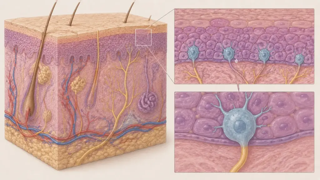
Merkel cell carcinoma is an aggressive form of skin cancer that develops from Merkel cells located in the basal layer of the epidermis, the outermost layer of the skin. These cells are neuroendocrine cells that play a role in the perception of touch and pressure. MCC is histopathologically characterized as a small, round, blue-cell tumor and is distinguished from other neuroendocrine cancers by perinuclear dot-like staining for cytokeratin-20 (CK20).
MCPyV is a major etiological factor in the development of MCC. Expression of the virus’s large tumor antigen (LT) and small tumor antigen (sT) disrupts cell cycle control by inactivating the pRb tumor suppressor protein. In UV-associated MCCs, TP53 and RB1 mutations are prominent. The incidence of MCC has tripled in recent years; this increase is attributed to both an aging population and improved diagnostic methods.
Assoc. Prof. Dr. Şengül emphasizes that in clinical practice, MCC is often misdiagnosed as a “simple skin infection” or a “sebaceous cyst,” leading to delayed diagnosis; she stresses that a rapidly growing, red-to-purple skin nodule requires immediate evaluation.
Symptoms of Merkel Cell Carcinoma
The symptoms of Merkel cell carcinoma vary depending on the tumor’s location and stage. They most commonly appear in areas exposed to the sun (face, head, neck, limbs):
- Rapidly growing skin nodule: A red, purple, or skin-colored, painless, firm mass; the average size at the time of diagnosis is 1–2 cm.
- Shiny surface: The skin overlying the tumor appears taut and shiny.
- Telangiectasia: Prominent superficial blood vessels.
- Ulceration: In advanced stages, the tumor surface may ulcerate.
- Regional lymph node involvement: Swelling in the neck, armpit, or groin; lymph node metastasis develops in 50% of cases.
- Signs of distant metastasis: Symptoms related to metastases in the liver, lungs, bones, or brain; distant metastasis is detected in 30% of cases.
Dermoscopic findings in Merkel cell carcinoma provide features supportive of the diagnosis. On dermoscopy, milky-red areas, atypical blood vessels, white amorphous areas, and cysts may be observed. However, dermoscopy is not diagnostic; a definitive diagnosis is established by biopsy and immunohistochemistry.
Treatment of Merkel Cell Carcinoma
Treatment for Merkel cell carcinoma is planned on a multidisciplinary basis according to the stage, tumor location, and the patient’s immune status:
Localized Disease (Stages I–II)
- Surgical Excision: Wide local excision or Mohs micrographic surgery; a 1–2 cm safety margin is targeted.
- Sentinel Lymph Node Biopsy (SLNB): Performed for staging in clinically node-negative patients; found to be positive in 30–40% of cases.
- Adjuvant Radiotherapy: Improves local control in high-risk patients (positive margins, lymphovascular invasion, large tumor); may reduce the recurrence rate by half.
- Adjuvant Immunotherapy: The ADMEC-O Phase II trial demonstrated that adjuvant nivolumab improved disease-free survival (DFS). The STAMP (pembrolizumab) and I-MAT (avelumab) Phase III trials are ongoing.
Advanced and Metastatic Disease (Stages III–IV)
- Immunotherapy (First-Line Treatment): Avelumab (JAVELIN Merkel 200) and pembrolizumab (KEYNOTE-017) are FDA-approved. In the KEYNOTE-017 trial, pembrolizumab achieved an ORR of %58, 3-year PFS of 39.1%, and 3-year OS of 59.4%. With avelumab, sustainable responses and long-term survival were observed during 4 years of follow-up.
- Nivolumab: In the CheckMate 358 trial, the ORR was 60%, median DOR was 60.6 months, median PFS was 21.3 months, and median OS was 80.7 months.
- Chemotherapy: A platinum-etoposide regimen is used in patients who are unresponsive to or contraindicated for immunotherapy; responses are short-lived and toxicity is high.
- Radiotherapy: For palliative pain and symptom control; stereotactic body radiotherapy (SBRT) may be considered in oligometastatic disease.
- Combination Therapies: Pembrolizumab + SBRT (NCT03304639) and the ipilimumab/nivolumab combination are being evaluated in refractory disease. The PANDORA Phase II trial is investigating the combination of pembrolizumab with platinum/etoposide.
| Treatment Agent | Study | ORR | Median OS | Status |
|---|---|---|---|---|
| Pembrolizumab | KEYNOTE-017 | 58% | 3 years: 59.4% | FDA-approved |
| Avelumab | JAVELIN Merkel 200 | 33% (refractory), 62% (1L) | ~20 months (refractory) | FDA-approved |
| Nivolumab | CheckMate 358 | 60% | 80.7 months | Clinical use |
| Platinum-etoposide | Historical data | 20–60% | <12 months | After immunotherapy |
According to Assoc. Prof. Dr. Nilay Şengül, immunotherapy has replaced chemotherapy as the standard first-line treatment for Merkel cell carcinoma; in particular, the durable responses achieved with pembrolizumab and avelumab have significantly altered the natural course of this aggressive cancer. However, the response rate to immunotherapy is low in immunocompromised patients (organ transplant recipients, HIV/AIDS); treatment decisions for this patient group should be made on a multidisciplinary basis.
Clinical Assessment by Assoc. Prof. Dr. Nilay Şengül
In my clinical practice, I frequently observe that most patients diagnosed with Merkel cell carcinoma have neglected the skin lesion, mistaking it for a “simple mole” or an “age spot.” Particularly in immunocompromised patients (organ transplant recipients, those on chronic steroid therapy), MCC follows a more aggressive course, and the prognosis worsens as diagnosis is delayed. According to Assoc. Prof. Dr. Nilay Şengül, a rapidly growing, painless, red-purple skin nodule must be evaluated via biopsy within 2–3 weeks; in MCC diagnosed at an early stage, the 5-year survival rate can reach 64% with surgery plus adjuvant radiation therapy. This approach highlights the vital importance of regular skin examinations, particularly in older patients and those who are immunocompromised.
From Diagnosis to Recovery: How Does the Process Proceed?
The diagnosis and treatment process for Merkel cell carcinoma progresses in stages depending on the patient’s clinical condition:
- Initial Examination and Evaluation: A detailed medical history is taken from patients presenting with suspected Merkel cell carcinoma. The growth rate of the lesion, pain status, skin color changes, and any lymph node swelling are assessed. A history of UV exposure, immunosuppression (organ transplantation, HIV/AIDS, chronic steroid use), and a family history of skin cancer are evaluated. During the physical examination, the size, color, borders, and mobility of the skin lesion are assessed, and regional lymph nodes are palpated.
- Tests and Imaging: A definitive diagnosis is established by incisional or puncture biopsy. In immunohistochemical staining, perinuclear dot-like staining for CK20, as well as positivity for synaptophysin and chromogranin A, are supportive findings. The viral status is evaluated via MCPyV immunohistochemistry or PCR. PET-CT or CT of the chest, abdomen, and pelvis is performed to screen for distant metastases; initial imaging results in upstaging in 12–20% of cases. SLNB is performed for staging in clinically node-negative patients.
- Treatment Planning: Findings are evaluated by a multidisciplinary oncology board (dermatology, surgical oncology, medical oncology, radiation oncology, pathology). For Stage I–II patients, surgical excision + SLNB + adjuvant radiation therapy (for high-risk cases) is planned. For Stage III–IV patients, immunotherapy (avelumab, pembrolizumab, or nivolumab) is the first-line treatment. In patients for whom immunotherapy is contraindicated, chemotherapy (platinum-etoposide) or palliative radiation therapy is considered.
- Surgical Procedure and Hospital Stay: Wide local excision or Mohs surgery is performed under local or general anesthesia. SLNB can be performed during the same session or as a separate procedure. The hospital stay lasts 1–3 days, depending on the extent of the surgery. Wound care and infection control are closely monitored. The decision regarding adjuvant therapy is made based on the pathology results.
- Post-Discharge Recovery: Wound healing after surgery is complete within 2–4 weeks. Adjuvant radiation therapy is initiated 4–6 weeks after surgery. Immunotherapy is administered intravenously every 2–3 weeks in cases of metastatic disease. As part of the follow-up plan, for the first 3 years (due to high risk of recurrence) and every 6 months thereafter. Immune-related adverse events (thyroiditis, hepatitis, pneumonitis, colitis) are closely monitored.
According to Assoc. Prof. Dr. Nilay Şengül, the risk of recurrence is high during the follow-up period after Merkel cell carcinoma treatment, particularly within the first 3 years; therefore, clinical and imaging follow-up every 3 months is critical. Additionally, the early diagnosis and management of immune-related adverse events in patients receiving immunotherapy are of vital importance for treatment continuity and quality of life.
Frequently Asked Questions
Is Merkel cell carcinoma contagious?
Merkel cell carcinoma itself is not contagious. However, the Merkel cell carcinoma virus (MCPyV), detected in 80% of cases, is widely transmitted through skin-to-skin contact; MCPyV antibodies are detected in 80% of adults. However, the viral infection is mostly asymptomatic, and only a very small proportion of cases progress to MCC. Immunosuppression is the primary factor that triggers the virus’s oncogenic potential.
Is Merkel cell carcinoma worse than melanoma?
Merkel cell carcinoma has a higher mortality rate than melanoma. The 5-year survival rate is 64% for Stage I–II MCC, whereas it is over 90% for melanoma at similar stages. MCC has a more aggressive course, metastasizes more rapidly, and is more resistant to chemotherapy. However, outcomes for MCC have improved dramatically with immunotherapy; sustainable responses achieved with pembrolizumab and avelumab have transformed the prognosis of this cancer.
How long is immunotherapy administered for Merkel cell carcinoma?
For metastatic Merkel cell carcinoma, pembrolizumab 2 mg/kg is administered every 3 weeks, avelumab 10 mg/kg every 2 weeks intravenously. Treatment continues until disease progression or unacceptable toxicity. In the KEYNOTE-017 trial, some patients received pembrolizumab for up to 2 years, and durable responses were observed. In adjuvant immunotherapy trials (STAMP, I-MAT), the treatment duration ranges from 6 to 12 months.
When does recurrence occur in Merkel cell carcinoma?
Recurrence in Merkel cell carcinoma is most common within the first 3 years after diagnosis. The local recurrence rate is between 40% and 50%. While the risk of recurrence is 5%–12% in patients with a negative sentinel lymph node biopsy (SLNB), this risk is higher in patients with a positive SLNB or those who underwent lymph node dissection. Regular follow-up is critical for early detection of recurrence; according to ESMO guidelines, follow-up every 3 months is recommended for the first 3 years.
Merkel cell carcinoma is a rare but extremely aggressive neuroendocrine skin cancer. MCPyV and UV exposure are the primary risk factors. High survival rates can be achieved with surgery plus adjuvant radiation therapy in the early stages. Immunotherapy (avelumab, pembrolizumab, nivolumab) has become the standard first-line treatment for metastatic disease and has dramatically improved outcomes. The diagnostic and treatment approach differs in immunocompromised patients; therefore, a multidisciplinary evaluation is of vital importance.
To evaluate your personal situation regarding Merkel cell carcinoma and have your questions answered, you can consult Assoc. Prof. Dr. Nilay Şengül. To schedule an appointment at the Memorial Şişli Hospital clinic, please call 444 7 888.
Sources
- PMC/NIH, “Immune Checkpoint Inhibitors in Merkel Cell Carcinoma of the Skin: A 2025 Comprehensive Review” — https://pmc.ncbi.nlm.nih.gov/articles/PMC12523286/
- PMC/NIH, “Merkel cell polyomavirus and associated Merkel cell carcinoma” — https://pmc.ncbi.nlm.nih.gov/articles/PMC8715208/
- PMC/NIH, “Merkel Cell Polyomavirus: A Newly Discovered Human Virus with Oncogenic Potential” — https://pmc.ncbi.nlm.nih.gov/articles/PMC3522868/
- NCCN Guidelines Version 2.2025, Merkel Cell Carcinoma — https://www.nccn.org/professionals/physician_gls/pdf/mcc.pdf
- ESMO Clinical Practice Guidelines, Merkel Cell Carcinoma — https://www.esmo.org/guidelines/guidelines-by-topic/rare-cancers/merkel-cell-carcinoma
- SEER/NCI, Cancer Statistics Facts: Merkel Cell Carcinoma — https://seer.cancer.gov/statfacts/html/merkel.html
- ClinicalTrials.gov, STAMP Trial (NCT03712605) — https://clinicaltrials.gov/study/NCT03712605