📌 What You Need to Know in a Nutshell
- Liposarcoma is the most common soft tissue sarcoma in adults; accounting for 15–20% of all soft tissue sarcomas, with an annual incidence of 2.5 per 1,000,000 people.
- The 5-year survival rate for well-differentiated liposarcoma is over 95%; for dedifferentiated liposarcoma, this rate drops to 50–70%.
- The extremities are the primary site in 52% of cases, and the retroperitoneum in 19%; the myxoid subtype is more common in patients under 50 years of age.
- R0 resection (negative surgical margins) is the most critical prognostic factor; achieving negative margins is difficult in retroperitoneal tumors.
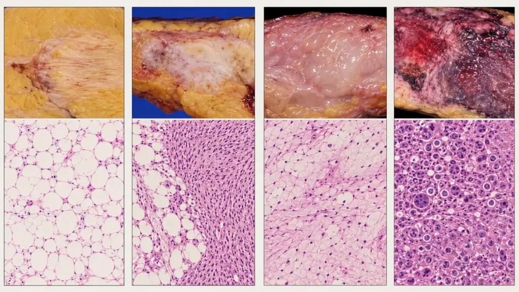
Liposarcoma is a malignant soft tissue tumor originating from adipose tissue. It is the most common type of sarcoma in adults, accounting for 15–20% of all soft tissue sarcomas. It is classified by the World Health Organization (WHO) into four main subtypes: well-differentiated, dedifferentiated, myxous, and pleomorphic. Each subtype has its own distinct molecular characteristics, clinical behavior, and treatment responses. Types of liposarcoma is critical for accurate diagnosis and personalized treatment.
What Is Liposarcoma?
Liposarcoma is a sarcoma that develops as a result of the malignant transformation of fat cells (adipocytes). It can occur in any anatomical region containing adipose tissue. It is most commonly found in the extremities (52%), the retroperitoneum (19%), the trunk (18%), and the head and neck region (9%). The annual incidence is approximately 2.5 cases per 1,000,000 people, and the peak incidence occurs between the ages of 50 and 65. There is a slight predominance in men compared to women (male-to-female ratio 1.2–1.5:1).
The molecular pathogenesis of liposarcoma varies depending on the subtype. In well-differentiated and dedifferentiated liposarcomas, MDM2 and CDK4 gene amplifications are detected; this leads to p53 pathway inactivation and cell cycle abnormalities. In myxous liposarcomas, FUS-DDIT3 or EWSR1-DDIT3 fusion genes are present. Pleomorphic liposarcoma, on the other hand, is characterized by complex chromosomal abnormalities.
What Is Dedifferentiated Liposarcoma?
Dedifferentiated liposarcoma (DDLPS) is an aggressive subtype that either arises from well-differentiated liposarcoma or develops de novo. The tumor exhibits an abrupt transition between areas of well-differentiated adipose tissue and areas of high-grade sarcoma. Retroperitoneal location is common. Dedifferentiation occurs in 10–15% of well-differentiated liposarcomas in retroperitoneal tumors and in less than 5% of those in extremity tumors. The 5-year survival rate is 50–70%; this rate is better in extremity tumors (55–70%) compared to retroperitoneal tumors.
Liposarcoma Symptoms
Symptoms of liposarcoma vary depending on the tumor’s location, size, and subtype. In the early stages, most patients are asymptomatic:
- Palpable mass: A mass in deep soft tissues that grows slowly or rapidly and is painless or causes mild pain; in retroperitoneal tumors, a sensation of abdominal distension.
- Pain: Occurs due to nerve or muscle compression; nighttime pain increases suspicion of malignancy.
- Muscle weakness and limited movement: In tumors of the extremities, especially those invading nerves or muscles.
- Retroperitoneal symptoms: Abdominal pain, weight loss, nausea/vomiting (gastrointestinal compression), leg edema (venous compression).
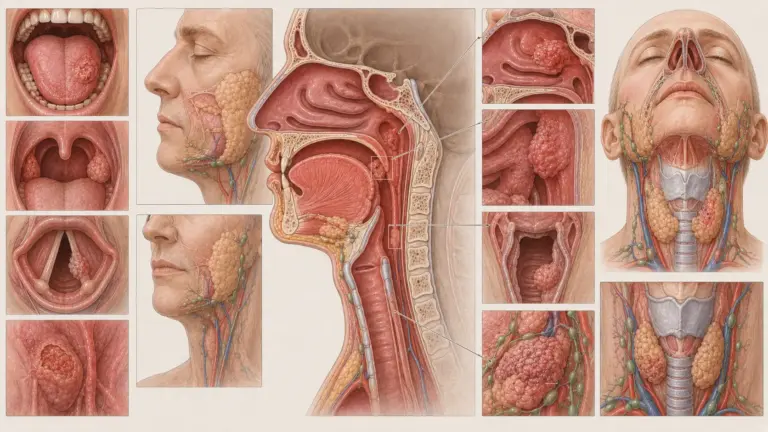
- Neck stiffness: Rare (9%); neck mass, difficulty swallowing, or shortness of breath.
Assoc. Prof. Dr. Şengül emphasizes that a significant proportion of patients diagnosed with liposarcoma in clinical practice mistakenly assume their mass is a “fat lump” (lipoma) and neglects it for a long time; she emphasizes that any deep, rapidly growing, or painful mass should be evaluated with imaging and a biopsy.
What Causes Liposarcoma?
The exact cause of liposarcoma is unknown. However, certain risk factors and molecular mechanisms have been identified:
- Genetic mutations: MDM2/CDK4 amplification (well-differentiated/dedifferentiated), FUS-DDIT3 fusion (mucinous), complex karyotype (pleomorphic).
- Radiation exposure: Secondary sarcoma development has been reported in areas previously treated with radiation therapy.
- Chronic lymphedema: Similar to the risk of angiosarcoma associated with Stewart-Treves syndrome, long-term lymphedema may increase the risk of liposarcoma.
- Chemical exposure: An increased risk of sarcoma has been reported in association with herbicides, chlorophenols, and vinyl chloride.
- Li-Fraumeni syndrome: Individuals who carry a TP53 mutation have an increased risk of soft tissue sarcoma.
According to Assoc. Prof. Dr. Nilay Şengül, most cases of liposarcoma are sporadic, and no clear risk factor can be identified; therefore, primary prevention strategies are limited. However, early evaluation of deep soft tissue masses significantly improves diagnostic and treatment outcomes.
Where Does Liposarcoma Occur?
Liposarcoma can occur in any area containing adipose tissue. Its anatomical distribution varies depending on the subtype:
- Extremities (52%): This is the most common site of occurrence, particularly the thigh and hip. Myxous and pleomorphic subtypes are more common.
- Retroperitoneum (19%): 70–80% of well-differentiated and dedifferentiated subtypes are retroperitoneal.
- Trunk (18%): Chest wall, abdominal wall, and pelvic region.
- Head and neck (9%): A rare site; liposarcoma in the neck is quite rare.
- Mediastinum and visceral organs (<%2): Primary liposarcoma of the lung is rare; liposarcoma of the lung typically indicates metastatic involvement.
Types of Liposarcoma
The four main subtypes of liposarcoma defined by the WHO exhibit different molecular characteristics, clinical behaviors, and treatment responses:
| Subtype | Incidence | Molecular Characteristics | 5-Year Survival |
|---|---|---|---|
| Well-Differentiated (WDLPS) | 40–45% | MDM2/CDK4 amplification | 95+% (with complete resection) |
| Undifferentiated (DDLPS) | 15–20% | MDM2/CDK4 amplification | 50–70% |
| Mixed (MLPS) | 30–35% | FUS-DDIT3 or EWSR1-DDIT3 fusion | 70–80% |
| Pleomorphic (PLPS) | 5–10% | Complex karyotype | 30–50% |
How Is Liposarcoma Treated?
Liposarcoma treatment is personalized based on the subtype, stage, location, and surgical margins:
- Surgical Resection: This is the gold standard treatment. The goal is R0 resection (negative microscopic margins). Wide local excision is performed for extremity tumors; compartment resection is performed for retroperitoneal tumors. The R0 margin is the most critical prognostic factor.
- Radiotherapy: It is used as neoadjuvant or adjuvant therapy. It improves local control in high-grade extremity tumors. In retroperitoneal sarcomas, preoperative radiation therapy may increase the chance of achieving a negative margin.
- Chemotherapy: Efficacy varies by subtype. Myxous liposarcomas are more sensitive to chemotherapy; a doxorubicin + ifosfamide regimen is used. Well-differentiated liposarcomas are resistant to chemotherapy. For metastatic diseasemay be treated with eribulin or trabectedin.
- Targeted Therapies: MDM2 inhibitors (idasanutlin, milademetan) and CDK4 inhibitors (palbociclib) are being evaluated in clinical trials. Pazopanib is a multi-target tyrosine kinase inhibitor used in advanced-stage soft tissue sarcomas.
- Immunotherapy: The response rate to immunotherapy in liposarcomas is low; agents such as pembrolizumab are being evaluated in limited subgroups.
According to Assoc. Prof. Dr. Nilay Şengül, it is critical that the initial surgery for liposarcoma be performed at an experienced sarcoma center; a failed or margin-positive initial surgery significantly increases the risk of recurrence and limits subsequent treatment options.
Liposarcoma Surgery
Liposarcoma surgery is performed using different techniques depending on the tumor’s location and size:
- Wide local excision: For tumors in the extremities, the tumor is removed along with surrounding healthy tissue. Efforts are made to preserve nerves and blood vessels.
- Compartment resection: In retroperitoneal tumors, the tumor, surrounding adipose tissue, and adjacent organs (if necessary) are removed.
- Amputation: Rarely; in extremity tumors where the nerve and vascular structures are completely invaded.
- Reconstruction: Reconstruction using a free flap or allograft may be necessary following extensive resection.
Post-Liposarcoma Surgery
The post-liposarcoma surgery period includes the recovery and follow-up phases. The hospital stay lasts 3–10 days, depending on the extent of the surgery. Postoperative pain management, wound care, and early mobilization are important.
Physical therapy following extremity surgery plays a critical role in restoring joint range of motion and muscle strength. Recovery of bowel function after retroperitoneal surgery may take several weeks. Adjuvant radiation therapy is initiated 4to 6 weeks after surgery. As part of the follow-up plan, local monitoring via MRI or CT is recommended every 3 months for the first 2 years and every 6 months for the following 3 years.
Clinical Assessment by Assoc. Prof. Dr. Nilay Şengül
In my clinical practice, I frequently observe the following: A significant proportion of patients diagnosed with liposarcoma have interpreted their mass as a “a fatty lump” and have delayed surgery. In particular, well-differentiated liposarcomas with a retroperitoneal location may not cause symptoms until they reach a diameter of 10–30 cm. According to Assoc. Prof. Dr. Nilay Şengül, a deep soft tissue mass must always be evaluated with magnetic resonance imaging (MRI) prior to biopsy; imaging directly influences the tumor’s margins, neurovascular relationships, and surgical planning. This approach optimizes both local control and functional outcomes, particularly by ensuring that the initial surgery is performed at an experienced sarcoma center.
From Diagnosis to Recovery: How Does the Process Proceed?
The liposarcoma diagnosis and treatment process proceeds in stages based on the patient’s clinical condition:
- Initial Examination and Evaluation: A detailed medical history is taken from patients presenting with suspected liposarcoma. The growth rate of the mass, the nature of the pain, weight loss, and current symptoms are assessed. A family history of sarcoma, prior radiation therapy, or the presence of chronic lymphedema is evaluated. During the physical examination, the mass’s size, mobility, firmness, skin changes, and regional lymph nodes are palpated.
- Tests and Imaging: Magnetic resonance imaging (MRI) is the gold standard for evaluating extremity tumors. CT is preferred for retroperitoneal tumors. Image-guided core needle biopsy is used for diagnosis and subtype determination; fine-needle aspiration (FNA) is insufficient. MDM2 FISH confirms the diagnosis of well-differentiated liposarcoma. Chest CT is performed to screen for lung metastases. PET-CT aids in staging high-grade tumors.
- Treatment Planning: Findings are evaluated in a multidisciplinary sarcoma board (orthopedic oncologist, surgical oncologist, medical oncologist, radiation oncologist, radiologist, pathologist) . For extremity tumors, wide local excision ± neoadjuvant/adjuvant radiation therapy is planned. For retroperitoneal tumors, compartment resection is the goal; preoperative radiation therapy may be considered. In metastatic disease, systemic therapy (chemotherapy, targeted therapies) is prioritized.
- Surgical Procedure and Hospital Stay: Liposarcoma surgery is performed under general anesthesia. The duration of surgery ranges from 2 to 6 hours, depending on the tumor’s size and location. The hospital stay lasts 3–10 days. Postoperative pain management, wound care, early mobilization, and physical therapy are initiated. The decision regarding adjuvant therapy is made based on pathology results.
- Recovery After Discharge: Full recovery occurs within 4–8 weeks. A return to light desk work is possible within 2–4 weeks. Strenuous physical activity and heavy lifting are restricted for the first 6–8 weeks. Physical therapy following extremity surgery should be initiated within 2–4 weeks. As part of the follow-up plan, local imaging (MRI/CT) and chest CT scans are recommended. In retroperitoneal tumors, the risk of local recurrence is between 40% and 60%; therefore, long-term follow-up is of vital importance.
According to Assoc. Prof. Dr. Nilay Şengül, long-term follow-up is critical during the post-treatment monitoring process for liposarcoma, particularly in well-differentiated liposarcomas with retroperitoneal location due to the risk of dedifferentiation; dedifferentiation can occur in 10–15% of tumors with retroperitoneal locations.
Frequently Asked Questions
What is a well-differentiated liposarcoma?
Well-differentiated liposarcoma (WDLPS) is the most common subtype of liposarcoma, accounting for 40-45% of cases. It is a slow-growing, low-grade tumor originating from mature adipocytes. It is characterized by MDM2 and CDK4 gene amplification. The 5-year survival rate after complete surgical resection is over 95%. However, in tumors with retroperitoneal, achieving negative margins is difficult; the local recurrence rate ranges from 40% to 60%. The recurrence rate is lower in extremity locations (10%–30).
Is liposarcoma dangerous?
Liposarcoma is a malignant tumor and can potentially lead to serious consequences. The degree of danger varies depending on the subtype: well-differentiated liposarcoma has a relatively good prognosis (95%+ 5-year survival rate), but the dedifferentiated and pleomorphic subtypes have an aggressive course. Retroperitoneal location indicates a poorer prognosis due to the difficulty in achieving negative margins. Early diagnosis and R0 surgical resection performed at an experienced center significantly improve outcomes.
Is liposarcoma cancer?
Yes, liposarcoma is cancer. It is a malignant (malignant) soft tissue sarcoma that originates from fat tissue. It should not be confused with a lipoma, which is a benign fat tumor. Liposarcoma can invade surrounding tissues, metastasize to distant organs, and be life-threatening if left untreated. A definitive diagnosis is made through biopsy and immunohistochemistry.
What is the most dangerous sarcoma?
The concept of the “most dangerous sarcoma” is relative; the prognosis varies depending on the subtype, stage, and location. Among liposarcoma subtypes, pleomorphic liposarcoma is the most aggressive; its 5-year survival rate ranges from 30% to 50%. Undifferentiated liposarcoma also has a poor prognosis. In general, among soft tissue sarcomas, angiosarcoma, pleomorphic liposarcoma, and undifferentiated liposarcoma are considered among the most aggressive types. However, each patient’s condition must be evaluated on an individual basis.
Where does liposarcoma metastasize?
The metastatic pattern of liposarcoma varies depending on the subtype. Pleomorphic liposarcoma tends to metastasize to the lungs (30–50% metastasis rate). Myxous liposarcoma, on the other hand, uniquely metastasizes to extrapulmonary sites; metastases to bone, soft tissue, the retroperitoneum, and the liver are common. Well-differentiated liposarcoma has minimal metastatic potential; however, it may metastasize following dedifferentiation. In dedifferentiated liposarcoma, the metastasis rate ranges from 15% to 30%; the lungs, liver, bone, and intra-abdominal regions may be involved.
Is liposarcoma fatal?
If left untreated or diagnosed at an advanced stage, liposarcoma can be life-threatening. However, many patients achieve long-term survival when diagnosed at an early stage and following complete surgical resection. The 5-year survival rate is over 95% for well-differentiated liposarcoma, 70–80% for myxous liposarcoma, 50–70% for dedifferentiated liposarcoma, and 30–50% for pleomorphic liposarcoma. Factors affecting prognosis include: subtype, stage, surgical margin status, location, and the presence of metastases.
What is the life expectancy for liposarcoma?
Life expectancy for liposarcoma varies significantly depending on the subtype. In well-differentiated liposarcoma, median survival can extend for years; the 5-year survival rate is over 95%. In myxous liposarcoma, the 5-year survival rate is between 70% and 80%. In dedifferentiated liposarcoma, the 5-year survival rate is between 50% and 70%, and in pleomorphic liposarcoma, it is between 30% and 50%. Retroperitoneal location, advanced age, high grade, and positive surgical margins worsen the prognosis. In metastatic myxous liposarcoma, the median disease-specific survival is 52.5 months.
How is liposarcoma surgery performed?
Liposarcoma surgery is is performed as either a wide local excision or a compartment resection, depending on the tumor’s location. In extremity tumors, the tumor and surrounding healthy tissue (typically 1–2 cm) are removed; efforts are made to preserve nerves and blood vessels. In retroperitoneal tumors, adjacent organs may also be removed. The surgery is performed under general anesthesia. The procedure lasts between 2 and 6 hours. The hospital stay is 3–10 days. The goal is an R0 resection (negative margins); this is the most important prognostic factor.
Does liposarcoma recur after removal?
The risk of liposarcoma recurrence varies depending on the subtype and location. In well-differentiated liposarcoma, the recurrence rate in the extremities is 10–30%, and in the retroperitoneum, it is 40–60%. In undifferentiated liposarcoma, the local recurrence rate is 20–30% in the extremities and 40–50%. The recurrence rate is lower in myxous liposarcomas. R0 resection significantly reduces the risk of recurrence. Regular follow-up is is critical for early detection of recurrence.
How long does liposarcoma surgery take?
The duration of liposarcoma surgery varies depending on the tumor’s size, location, and the extent of the surgery. For tumors in the extremities, wide local excision typically takes 2–4 hours. For retroperitoneal tumors, if compartment resection and removal of adjacent organs are required, the surgery may take up to 4to 6 hours. In complex cases requiring nerve and vascular reconstruction, the duration may be even longer. The duration of surgery is also related to the surgeon’s experience and the volume of cases at the center.
Liposarcoma is the most common soft tissue sarcoma in adults and is classified into four main subtypes (well-differentiated, dedifferentiated, myxous, and pleomorphic). Each subtype has its own distinct molecular characteristics, clinical behavior, and treatment responses. R0 surgical resection is the most critical prognostic factor across all subtypes. Early diagnosis, initial surgery performed at an experienced sarcoma center, and a multidisciplinary treatment approach directly impact survival and quality of life. Regular follow-up is of vital importance, particularly for tumors located in the retroperitoneum.
To discuss your specific situation regarding liposarcoma and have your questions answered, you can consult with Assoc. Prof. Dr. Nilay Şengül. To schedule an appointment at the Memorial Şişli Hospital clinic, please call 444 7 888.
Sources
- PMC/NIH, “Liposarcoma – StatPearls” — https://www.ncbi.nlm.nih.gov/books/NBK538265/
- PMC/NIH, “Recent advances in the management of liposarcoma” — https://pmc.ncbi.nlm.nih.gov/articles/PMC5224678/
- Nature/Scientific Reports, “Survival and prognostic factors among different types of liposarcomas based on the SEER database” — https://www.nature.com/articles/s41598-025-85937-9
- PMC/NIH, “Prognostic factors of metastatic myxoid liposarcoma” — https://pmc.ncbi.nlm.nih.gov/articles/PMC7491192/
- NCCN Guidelines, Soft Tissue Sarcoma — https://www.nccn.org/professionals/physician_gls/pdf/sarcoma.pdf
- ESMO Guidelines, Soft Tissue and Visceral Sarcomas — https://www.esmo.org/guidelines/guidelines-by-topic/sarcoma-and-gist