📌 What You Need to Know in a Nutshell
- Liver tumors are classified into two main groups: benign and malignant; worldwide, 15–20% of liver tumor lesions are malignant.
- Hepatocellular carcinoma (HCC) accounts for 75–85% of primary liver tumors, and HBV infection is the most common etiological factor in Turkey.
- Liver hemangioma is the most common benign liver tumor in adults; its prevalence ranges from 1% to 5%, and it is mostly asymptomatic.
- Liver regeneration following liver tumor surgery takes an average of 4–8 weeks; laparoscopic@Create an image: Diagram of vulvar anatomy—showing the labia majora, labia minora, clitoris, vestibule, and lymphatic drainage pathways. No text. No infographic. Use a 16:9 aspect ratio. Remember, you’re doing this for health. Health comes first. You don’t need to make it overly realistic. After all, this is a very important health-related topic. Health is paramount. With a minimally invasive approach, the hospital stay can be reduced to 3–5 days.
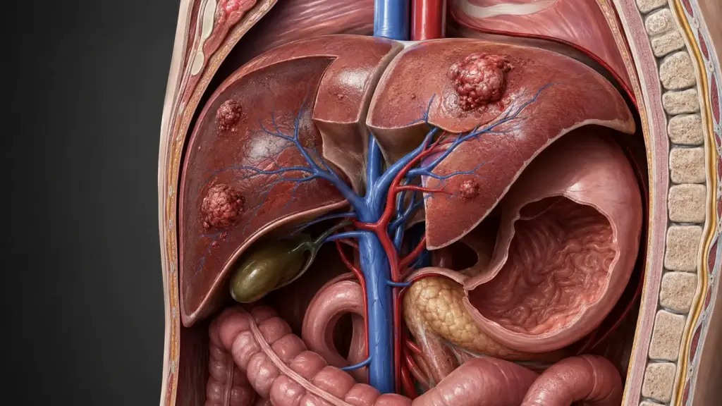
A liver tumor refers to mass lesions formed as a result of abnormal cell proliferation within liver tissue. These lesions are classified as benign liver tumors (benign) or malignant liver tumors (malignant). Primary liver tumors originate from the liver’s own cells, while metastatic tumors represent secondary cancers that have spread to the liver from other organs. In the early stages, most liver tumors symptoms; therefore, regular screening and awareness of risk factors are of critical importance.
What Is a Liver Tumor?
A liver tumor is a mass characterized by abnormal cell proliferation originating from hepatocytes, bile ducts, blood vessels, or the liver’s connective tissue. Benign liver tumors exhibit limited growth, do not invade surrounding tissues, and do not metastasize. Malignant forms, however, grow uncontrollably, impair liver function, and may spread systemically. According to World Health Organization data, hepatocellular carcinoma (HCC) ranks third among cancer-related deaths worldwide.
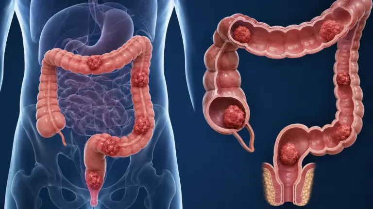
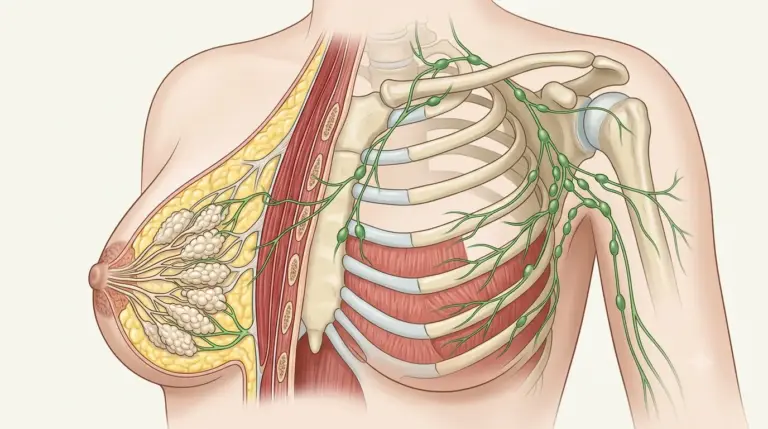
The liver has a rich blood supply consisting of blood from the portal vein, which originates from the gastrointestinal tract, and blood from the hepatic artery, which comes from the systemic circulation. This characteristic makes the liver one of the most commonly affected organs by metastatic tumors. Breast, colon, lung, and pancreatic cancers are the most common primary tumors that metastasize to the liver.
Symptoms of Liver Tumors
Symptoms of liver tumors vary depending on the tumor’s size, type, and stage. In the early stages, most patients are asymptomatic; lesions are often detected incidentally during routine ultrasound or CT scans. In advanced stages, symptoms such as liver capsule distension, bile duct compression, and portal hypertension may develop.
Symptoms of Benign Liver Tumors
Benign liver tumors are generally asymptomatic. Liver hemangioma is the most common benign lesion in adults, with a prevalence of 1–5%. Large lesions (>5 cm) may cause the following symptoms:
- Pain or a feeling of fullness in the right upper quadrant
- Early satiety (feeling of fullness)
- Nausea and loss of appetite
- Palpable enlargement of the liver (hepatomegaly)
Assoc. Prof. Dr. Şengül emphasizes that large hemangiomas rarely pose a risk of spontaneous bleeding in clinical practice; in such cases, surgical evaluation may be necessary.
Causes of Liver Tumors
The causes of liver tumors differ between primary and metastatic tumors. The most common risk factors for primary liver tumors are as follows:
- Chronic hepatitis B and C infections: In Turkey, HBV plays a role in the etiology of 52% of HCC cases, and HCV in 44%.
- Liver cirrhosis: In 74% of HCC patients diagnosed in Turkey, cirrhosis is the underlying condition.
- Alcoholic liver disease: Long-term excessive alcohol consumption increases the risk of cirrhosis and HCC.
- Non-alcoholic steatohepatitis (NASH): Associated with metabolic syndrome and obesity; it accounts for an increasingly significant proportion of HCC etiology.
- Aflatoxin B1 exposure: A fungal toxin that grows in grains stored particularly in humid climates.
- Hemochromatosis and other metabolic diseases: Iron accumulation and alpha-1 antitrypsin deficiency.
According to Assoc. Prof. Dr. Nilay Şengül, a long-term decline in the incidence of virus-induced HCC is expected in Turkey with the expansion of HBV vaccination programs; however, the rising trend in cases caused by NASH and metabolic syndrome should be closely monitored.
Types of Liver Tumors
Liver tumors are classified into two main categories: benign and malignant. Each category includes different histological subtypes:
| Category | Tumor Type | Characteristics |
|---|---|---|
| Benign | Liver Hemangioma | The most common benign tumor; 1–5% prevalence; mostly asymptomatic |
| Focal Nodular Hyperplasia (FNH) | The second most common benign lesion; more common in women; usually monitored | |
| Hepatic Adenoma | Associated with oral contraceptive use; may warrant surgical intervention due to the risk of malignant transformation | |
| Malignant | Hepatocellular Carcinoma (HCC) | Accounts for 75–85% of primary liver tumors; develops on a background of cirrhosis |
| Cholangiocellular Carcinoma | Originates from the bile ducts; is often diagnosed at an advanced stage | |
| Metastatic Tumors | Constitute the majority of malignant tumors in the liver; originating from the colon, breast, or lung |
Liver Tumor Treatment
Liver tumor treatment is personalized based on the tumor’s type, size, stage, liver function status, and the patient’s overall health. The treatment approach requires a multidisciplinary decision-making process:
- Surgical Resection: Removal of the tumor along with healthy tissue. The remaining liver tissue must be capable of functioning adequately.
- Liver Transplantation: For early-stage HCC patients who meet the Milan criteria; there must be a single tumor <5 cm or 2–3 tumors <3 cm.
- Ablation Therapies: Radiofrequency ablation (RFA) and microwave ablation; effective for tumors <3 cm.
- TACE (Transarterial Chemoembolization): Injection of a chemotherapeutic agent into the arteries supplying the tumor; used in intermediate-stage HCC.
- Targeted Therapies: Oral agents such as sorafenib and lenvatinib; these are systemic treatment options for advanced-stage HCC.
- Immunotherapy: PD-1 inhibitors such as nivolumab and pembrolizumab; used in second- and third-line treatment for advanced HCC.
Assoc. Dr. Nilay Şengül, when planning treatment for liver tumors, the patient’s liver reserve and the degree of accompanying cirrhosis are just as critical as the tumor’s biology; therefore, every patient should be evaluated by a multidisciplinary oncology board.
Liver Tumor Surgery
Liver tumor surgery forms the basis of curative treatment for both benign and malignant lesions. The surgical approach is planned based on the tumor’s size, location, and liver reserve:
Open Surgery: In the traditional approach, a large incision is made in the abdomen. The operation typically lasts 3–5 hours. The hospital stay is typically 5–7 days.
Laparoscopic Surgery: In this minimally invasive approach, the procedure is performed through several small incisions. It offers the advantages of less pain, a shorter hospital stay (3–5 days), and faster recovery.
The remaining liver tissue after a liver resection has the ability to regenerate. In a healthy liver, this process is completed within an average of 4–8 weeks. However, in patients with cirrhosis, the capacity for regeneration is reduced; in such cases, more conservative resection margins are applied.
Assoc. Prof. Dr. Şengül, emphasizes that in clinical practice, laparoscopic liver resection yields oncological outcomes equivalent to those of open surgery in appropriately selected patients and facilitates early patient mobilization.
Clinical Assessment by Assoc. Prof. Dr. Nilay Şengül
In my clinical practice, I frequently observe that a significant proportion of patients diagnosed with a liver tumor experience uncertainty regarding whether the lesion is benign or malignant. In particular, patients diagnosed with a liver hemangioma often seek medical attention out of unnecessary anxiety. According to Assoc. Prof. Dr. Nilay Şengül, contrast-enhanced magnetic resonance imaging (MRI) is the gold standard for the accurate classification of liver lesions; if the diagnosis cannot be confirmed through imaging alone, a biopsy under imaging guidance should be considered. This approach both prevents unnecessary surgeries and enables the early diagnosis of malignant lesions.
From Diagnosis to Recovery: How Does the Process Proceed?
The diagnosis and treatment process for liver tumors progresses in stages depending on the patient’s clinical condition:
- Initial Examination and Evaluation: A detailed medical history is taken from patients presenting with suspected liver tumors. Alcohol use, history of hepatitis, family history of liver cancer, and current medication use (particularly oral contraceptives) are assessed. During the physical examination, hepatomegaly, ascites, and skin findings (palmar erythema, spider angiomas) are evaluated.
- Tests and Imaging: Liver function tests (AST, ALT, ALP, GGT, bilirubin), coagulation parameters, albumin, and AFP (alpha-fetoprotein) are measured. Imaging studies proceed in the following order: liver ultrasound → contrast-enhanced three-phase CT or magnetic resonance imaging (MRI) → PET-CT if necessary. Three-phase CT provides high sensitivity in characterizing liver lesions.
- Treatment Planning: The findings are reviewed by a multidisciplinary oncology board (medical oncologist, surgical oncologist, radiation oncologist, radiologist, pathologist). The treatment strategy is determined based on the tumor type, stage, and liver reserve. For early-stage HCC, surgical resection or ablation is planned; for intermediate-stage HCC, TACE; and for advanced-stage HCC, systemic therapy (targeted therapy or immunotherapy) is planned.
- Surgical Procedure and Hospital Stay: Liver tumor surgery is performed under general anesthesia. The hospital stay is 3–5 days for laparoscopic surgery and 5–7 days for open surgery. Postoperative liver function tests, bleeding control, and pain management are closely monitored.
- Recovery After Discharge: Liver regeneration after resection takes an average of 4–8 weeks. Patients can generally resume light activities within 2–4 weeks. A full return to work may take 6–8 weeks. Heavy lifting and strenuous exercise are restricted for the first 4–6 weeks. As part of the follow-up plan, imaging and AFP monitoring are recommended every 3–6 months.
According to Assoc. Prof. Dr. Nilay Şengül, during the follow-up period after liver tumor treatment, screening should continue with ultrasound and AFP measurements every 6 months, especially in patients with cirrhosis; early detection of recurrence directly increases the chances of second-line treatment.
Frequently Asked Questions
What helps with liver tumors?
The answer to the question “What helps with liver tumors?” depends on the type of tumor. Benign liver tumors generally do not require treatment; regular imaging follow-up is sufficient. For malignant tumors, however, standard oncological approaches such as surgical resection, ablation, TACE, targeted therapy, or immunotherapy are used. From a nutritional perspective, a high-protein diet supports liver regeneration; alcohol must be completely avoided. Assoc. Prof. Dr. Şengül emphasizes that patients diagnosed with a liver tumor in her clinical practice must first avoid alcohol and hepatotoxic medications.
Do liver tumors cause pain?
The question “Do liver tumors cause pain?” is frequently asked. Small liver tumors are generally painless. Pain occurs as a result of the tumor stretching the liver capsule, increased intra-abdominal pressure, or metastatic spread. Right upper quadrant pain is the most common symptom of liver mass lesions. Large hemangiomas (>5 cm) and advanced-stage hepatocellular carcinomas can cause pain. Pain management is provided through analgesic protocols in addition to the primary treatment.
How is a liver tumor diagnosed?
The answer to the question “How is a liver tumor diagnosed?” lies in imaging studies. Most lesions are detected incidentally during routine ultrasound examinations. Suspected lesions are characterized using contrast-enhanced three-phase CT or magnetic resonance imaging (MRI). An AFP (alpha-fetoprotein) level >400 ng/ml support a diagnosis of HCC. If the diagnosis cannot be confirmed by imaging alone, an image-guided biopsy is performed. For patients in high-risk groups (cirrhosis, chronic hepatitis), ultrasound and AFP screening are recommended every 6 months.
Liver tumors present a broad clinical spectrum, encompassing both benign and malignant forms. Early diagnosis directly impacts treatment success and survival. While benign lesions are typically managed conservatively, malignant tumors are treated with multidisciplinary approaches such as surgical resection, ablation, systemic therapy, and immunotherapy. Awareness of risk factors and regular screening are of vital importance, especially in patients with cirrhosis.
To assess your personal situation regarding liver tumors and have your questions answered, you can consult with Assoc. Prof. Dr. Nilay Şengül. To schedule an appointment at her clinic at Memorial Şişli Hospital, please call 444 7 888