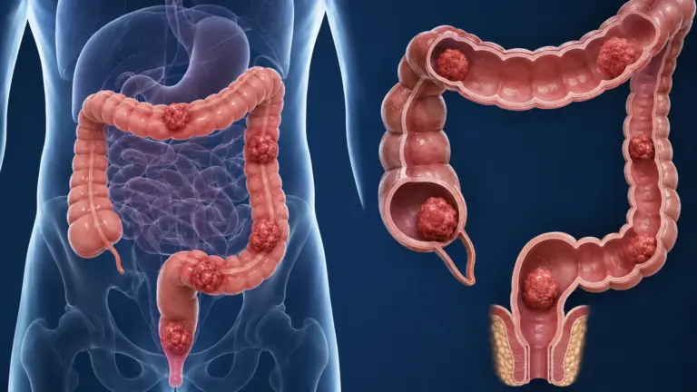

📌 What You Need to Know in a Nutshell
- Kidney cancer develops when cells in the kidney tissue multiply uncontrollably; the most common subtype is clear cell renal cell carcinoma (70–80%).
- In early stages (Stages I–II), the 5-year survival rate is 85–95%; curative results can be achieved with surgical treatment.
- Immune checkpoint inhibitors and targeted therapies can extend median survival to 40–50 months in metastatic disease.
- Smoking, obesity, and high blood pressure are the most significant modifiable risk factors for kidney cancer.
Kidney cancer is a malignant tumor originating from the tubular epithelial cells of the kidneys. Approximately 434,000 new cases are diagnosed worldwide each year, and it is about twice as common in men than in women. The disease typically presents between the ages of 55 and 74; because it does not cause symptoms in the early stages, it is frequently detected incidentally during radiological examinations.
[VISUAL SUGGESTION: An anatomical diagram showing kidney anatomy and the site of renal cell carcinoma — Alt text: “Diagram of kidney anatomy and the site of renal cell carcinoma”]
What Is Kidney Cancer?
Kidney cancer is a malignant neoplasm resulting from the uncontrolled growth of cells in the renal parenchyma. Approximately 90% of cases are of the renal cell carcinoma (RCC) subtype; the vast majority of these are clear cell RCC (70–80%). Other subtypes include papillary RCC (10–15%), chromophobe RCC (5%), and collecting duct carcinoma (<1%). Urothelial carcinomas originating in the renal pelvis, however, have a different biology than RCC.
Renal cell carcinoma is the most common cancer associated with a von Hippel-Lindau (VHL) gene mutation. The VHL gene mediates the degradation of the hypoxia-responsive factor HIF-1α; a mutation in this gene leads to the accumulation of HIF-1α, which in turn drives angiogenesis and tumor growth. This mechanism has formed the basis for the development of targeted therapies.
Symptoms of Kidney Cancer
Symptoms of kidney cancer are often absent in the early stages. Approximately 50–60% of patients are asymptomatic at the time of diagnosis and are detected incidentally during imaging studies. In patients who do exhibit symptoms, the classic triad—gross hematuria (blood in the urine), flank pain (pain in the lower back), and a palpable mass—is present in only 10% of patients.
Common symptoms include:
- Gross hematuria (visible blood in the urine): This results from the tumor’s spread to the collecting system.
- Flank pain: Associated with capsular stretching or invasion of perirenal structures by the tumor.
- Intentional weight loss: Loss of more than 5% of body weight in the past 6 months.
- Fatigue and anemia: Associated with chronic blood loss or paraneoplastic syndromes.
- Fever: Paraneoplastic fever is defined as a non-infectious, low-grade fever.
Paraneoplastic syndromes also provide important clues in the diagnosis of kidney cancer. Erythrocytosis (hematocrit >50%), hypercalcemia (serum calcium >10.5 mg/dL), hypertension (increased renin secretion), and Stauffer syndrome (a triad of liver dysfunction, fever, and anemia) are the most common.
Symptoms of Adrenal Cancer
The symptoms of adrenal gland cancer present a different clinical picture than those of kidney cancer. Adrenal tumors usually cause symptoms because they are hormonally active. In cortisol-secreting tumors, Cushing’s syndrome (central obesity, facial fullness, purple striae, hypertension); Conn’s syndrome (hypertension, hypokalemia, muscle weakness) in aldosterone-secreting tumors; and the triad of paroxysmal hypertension, sweating, headache, and tachycardia in catecholamine-secreting pheochromocytomas. Hormone-inactive adrenal tumors are generally asymptomatic and are detected through imaging.
According to Assoc. Prof. Dr. Nilay Şengül, distinguishing between adrenal cancer and kidney cancer is of critical importance in clinical practice; the hormonal profile and imaging characteristics of adrenal tumors (Hounsfield units <10, rapid contrast washout) are key in the differential diagnosis.
What Causes Kidney Cancer?
Both genetic and environmental factors play a role in the development of kidney cancer. The most significant modifiable risk factor for the disease is smoking; the risk is 1.5 to 2.5 times higher in smokers than in nonsmokers. When smoking is quit, the risk decreases over time but does not return to the level of a person who has never smoked.
Risk Factors Table:
| Risk Factor | Increase in Risk | Mechanism |
|---|---|---|
| Smoking | 1.5–2.5 times | N-nitroso compounds, cadmium accumulation |
| Obesity (BMI >30) | 1.5–2 times | Insulin resistance, adipokine dysregulation |
| High blood pressure | 1.5–2 times | Chronic hypoxic damage, activation of the renin-angiotensin system |
| Family history | 2–4 times | VHL, MET, and BAP1 gene mutations |
| Long-term dialysis | 3–5 times | Cystic kidney disease, acquired cystic disease |
| Asbestos exposure | 1.4–2 times | Chronic inflammation, DNA damage |
Hereditary kidney cancer syndromes include von Hippel-Lindau (VHL), hereditary papillary RCC (MET mutation), Birt-Hogg-Dubé (FLCN mutation), and hereditary leiomyomatosis-associated RCC (FH mutation). In these syndromes, patients’ lifetime risk of kidney cancer ranges from 30% to 70%.
Stages of Kidney Cancer
Kidney cancer stages are determined according to the AJCC TNM 8th Edition classification. Staging is based on tumor size (T), lymph node involvement (N), and the presence of distant metastases (M). The stage is the most important factor in both treatment planning and determining prognosis.
Table of 5-year survival rates by stage:
| Stage | TNM Definition | 5-Year Survival (%) | Treatment Approach |
|---|---|---|---|
| Stage 1 | T1 (≤7 cm), N0, M0 | >90–95% | Partial nephrectomy (preferred) |
| Stage 2 | T2 (>7 cm, confined to the kidney), N0, M0 | 75–85% | Radical nephrectomy |
| Stage 3 | T3 (perirenal fat/venous invasion), N0–1, M0 | 50–70% | Radical nephrectomy ± adjuvant immunotherapy |
| Stage 4 | T4 or N2 or M1 (metastatic) | 10–20% | Systemic therapy (TKI + ICI combination) |
Stage 1 Kidney Cancer
Stage 1 kidney cancer is defined as a tumor that is smaller than 7 cm and confined to the kidney. The 5-year survival rate for this stage is over 90%. Partial nephrectomy (removal of only the tumor-containing portion of the kidney) is the preferred treatment; this approach preserves kidney function. For T1a (<4 cm) tumors, laparoscopic or robotic partial nephrectomy is the standard treatment. For T1b (4–7 cm) tumors, partial nephrectomy is preferred if technically feasible; if not, radical nephrectomy may be performed.
Stage 2 Kidney Cancer
Stage 2 kidney cancer refers to a tumor that is larger than 7 cm but is confined to the kidney. The 5-year survival rate at this stage is between 75% and 85%. Radical nephrectomy (removal of the entire kidney) is the standard treatment approach. It is divided into two subtypes: T2a (7–10 cm) and T2b (>10 cm). In T2 tumors, postoperative survival is high as long as there is no lymph node involvement; adjuvant therapy is generally not recommended.
Stage 3 Kidney Cancer
Stage 3 kidney cancer involves spread to the perirenal fat, the renal vein, or a single regional lymph node. The 5-year survival rate for this stage is between 50% and 70%. According to the KEYNOTE-564 study, adjuvant pembrolizumab treatment in high-risk Stage III patients significantly prolonged progression-free survival. Planning for systemic therapy after surgery is based on the pathological stage and risk scoring (Leibovich score).
Stage 4 Kidney Cancer
Stage 4 kidney cancer (metastatic disease) refers to the spread of the tumor beyond the kidney to distant organs. At this stage, survival in Stage 4 kidney cancer varies depending on treatment response and metastatic burden; generally, the 5-year survival rate ranges from 10% to 20%. In recent years, median survival has been extended to 40–50 months with combinations of immune checkpoint inhibitors (nivolumab, pembrolizumab) and tyrosine kinase inhibitors (axitinib, lenvatinib). Symptoms experienced in the advanced stages of kidney cancer include fatigue, weight loss, bone pain, and shortness of breath (if there are lung metastases).
Assoc. Prof. Dr. Şengül emphasizes that in her clinical practice, median survival has increased dramatically over the past 5 years with combination therapy of immunotherapy and TKIs in patients with metastatic kidney cancer. In particular, long-term complete response rates have been reported with these combination therapies in patients classified as low-risk according to the IMDC (International Metastatic RCC Database Consortium).
Kidney Cancer Treatment
Kidney cancer treatment is personalized based on the stage, tumor size, histological subtype, and the patient’s overall condition. Treatment approaches are categorized into two main groups: localized disease (Stages I–III) and metastatic disease (Stage IV).
Treatment of Localized Disease:
- Surgical treatment: This forms the foundation of kidney cancer treatment. Options include partial nephrectomy (organ-preserving) or radical nephrectomy.
- Ablative therapies: Radiofrequency ablation (RFA) and cryotherapy offer alternatives for T1a (<3 cm) tumors and for patients at high surgical risk.
- Active Surveillance: This may be preferred for T1a (<2 cm) tumors, as well as for patients at high surgical risk, the elderly, and those with comorbidities. The average annual growth rate has been reported as 0.3 cm.
Treatment of Metastatic Disease:
- Immune checkpoint inhibitors (ICI): The combination of nivolumab (PD-1 inhibitor) and ipilimumab (CTLA-4 inhibitor) is the standard first–line treatment for patients with intermediate-to-high IMDC risk.
- TKI + ICI Combinations: Pembrolizumab + axitinib, nivolumab + cabozantinib, and pembrolizumab + lenvatinib are FDA-approved combinations.
- TKI monotherapy: Agents such as sunitinib, pazopanib, and cabozantinib are used in patients who are ineligible for or have contraindications to ICI therapy.
- Cytoreductive nephrectomy: Removal of the primary tumor in selected metastatic patients may prolong survival when combined with systemic therapy.
Treatment comparison table:
| Treatment Method | Indication | Median Survival | Side Effects |
|---|---|---|---|
| Partial Nephrectomy | T1a-T1b, single kidney | >120 months | Bleeding, urinary fistula, decreased GFR |
| Radical Nephrectomy | T2–T3, large tumor | 100–120 months | Risk of chronic kidney disease |
| Nivo + Ipi | Metastatic, IMDC intermediate-high | 47.6 months | Hepatitis, colitis, hypophysitis, pneumonitis |
| Pembro + Axi | Metastatic, all risk groups | 45.7 months | Hypertension, hepatotoxicity, thyroiditis |
| Sunitinib | Metastatic, ICI contraindicated | 26.4 months | Hypertension, palmar-plantar erythrodysesthesia, myelosuppression |
Kidney Cancer Surgery
Kidney cancer surgery is the primary treatment for localized disease. The type of surgery is determined based on tumor size, location, the patient’s kidney function, and comorbidities.
Partial Nephrectomy (Nephron-Sparing Surgery):
Only the tumor-containing portion of the kidney is removed, while the healthy parenchyma is preserved. This is the standard treatment for T1a (<4 cm) tumors. It is also the preferred option for T1b (4–7 cm) tumors if technically feasible. Robotic surgery, 3D imaging, and intraoperative ultrasound ensure precise resection. The goal is to keep ischemia time under 25 minutes; exceeding this duration increases the risk of permanent kidney damage.
Radical Nephrectomy:
The entire kidney, perirenal fat, Gerota’s fascia, and regional lymph nodes are removed. It is performed in T2 (>7 cm) and higher stages, in the presence of venous invasion, or when partial nephrectomy is not technically feasible. Laparoscopic and robotic approaches result in less bleeding, a shorter hospital stay, and faster recovery compared to open surgery.
Comparison of Surgical Approaches:
| Approach | Hospital Stay | Blood Loss | Pain Score | Return to Work |
|---|---|---|---|---|
| Open Surgery | 5–7 days | High | High | 6–8 weeks |
| Laparoscopic | 2–4 days | Moderate | Moderate | 3–4 weeks |
| Robotic Surgery | 1–3 days | Low | Low | 2–3 weeks |
According to Assoc. Prof. Dr. Nilay Şengül, robotic partial nephrectomy offers superior results in terms of surgical margin safety and preservation of kidney function, particularly in cases of complex-located and hilar tumors. 3D imaging and Firefly fluorescence technology enable precise identification of vascular structures and tumor margins.
Clinical Assessment by Assoc. Prof. Dr. Nilay Şengül
In my clinical practice, I frequently observe the following: Approximately 40% of patients diagnosed with kidney cancer are incidentally detected during an abdominal ultrasound or CT scan performed for another reason. This situation increases the likelihood of detecting the disease at an early stage and improves the chances of curative surgery. However, in symptomatic patients, the diagnosis is usually made at more advanced stages.
According to Assoc. Prof. Dr. Nilay Şengül, genetic profiling is becoming increasingly important in kidney cancer. The presence of VHL, PBRM1, BAP1, and SETD2 mutations serves as a guide for both prognosis and treatment selection. In particular, patients with BAP1 mutations tend to have a more aggressive course of the disease and respond better to immunotherapy. For this reason, comprehensive molecular analysis is recommended for all patients with metastatic disease.
Assoc. Prof. Dr. Şengül emphasizes that, in her clinical practice, the use of adjuvant pembrolizumab significantly prolongs progression-free survival in high-risk Stage III patients. Based on the results of the KEYNOTE-564 study, a 1-year adjuvant treatment regimen is planned for this patient group.
From Diagnosis to Recovery: How Does the Process Proceed?
- Initial Examination and Evaluation: A detailed medical history is taken from the patient; smoking history, family history, previous kidney diseases, and current symptoms are recorded. During the physical examination, palpation of an abdominal mass, lymph node examination, and vital signs are assessed. Laboratory tests include a complete blood count, biochemistry (creatinine, GFR, electrolytes), urinalysis, and liver function tests.
- Imaging Studies: Contrast-enhanced abdominal CT (triphasic protocol) is the standard diagnostic method; tumor size, location, venous invasion, and lymph node status are evaluated. A chest CT is ordered to assess for lung metastases, and a bone scan or PET-CT is ordered if bone metastases are suspected. A brain MRI is performed if neurological symptoms are present or in high-risk patients.
- Surgical Process: Preoperative anesthesia evaluation, blood typing, and infection screening are performed. The type of surgery (partial or radical nephrectomy) and approach (open, laparoscopic, or robotic) are determined. The surgery typically lasts 2–4 hours. In partial nephrectomy, the goal is to keep ischemia time <25 minutes.
- Hospital Stay and Discharge: A hospital stay of 1–3 days is expected after robotic surgery, 2–4 days after laparoscopic surgery, and 5–7 days after open surgery. Prior to discharge, the drain is removed, oral intake is initiated, and pain management is ensured.
- Post-Discharge Recovery: Light activity is recommended for the first 2 weeks; heavy lifting (>5 kg) and exercise are restricted for 4–6 weeks. Return to work varies between 2 and 6 weeks, depending on the surgical approach. The need for adjuvant therapy is determined based on the pathology results. Follow-up schedule: abdominal CT and chest X-ray every 6 months for the first 2 years, then annually for the next 3 years.
Blood Test Results for Kidney Cancer
Blood test results in kidney cancer provide important clues for both diagnosis and follow-up. Laboratory findings are used to assess paraneoplastic syndromes, kidney function, and overall health status.
Key laboratory parameters:
- Serum creatinine and GFR: These assess kidney function. In patients scheduled for partial nephrectomy, a preoperative GFR >60 mL/min/1.73 m² is preferred.
- Complete blood count: Anemia (Hb <12 g/dL in men, <11 g/dL in women), erythrocytosis (hematocrit >50%), or thrombocytosis (>400,000/μL) may be paraneoplastic findings.
- Serum calcium: Hypercalcemia (>10.5 mg/dL) is seen in tumors that secrete PTHrP.
- LDH: Elevated LDH (>1.5 times the upper limit of normal) is a prognostic factor in IMDC risk scoring.
- Albumin: Low albumin (<3.5 g/dL) is a poor prognostic marker in the IMDC scoring system.
- CRP: Elevated CRP is associated with systemic inflammation and a poor prognosis.
Can kidney cancer be detected through blood tests? Kidney cancer cannot be diagnosed directly through a single blood test. However, LDH, calcium, complete blood count, and kidney function tests are helpful in the diagnostic process. Currently, circulating tumor DNA (ctDNA) analyses are in the research phase for monitoring treatment response and residual disease in metastatic disease.
Frequently Asked Questions
Can you get cancer in the kidney?
Yes, cancer can develop in the kidney. Kidney cancer is a malignant tumor resulting from the uncontrolled proliferation of cells in kidney tissue. Approximately 434,000 new cases are diagnosed worldwide each year. The most common type is renal cell carcinoma (90%).
How does kidney cancer develop?
Kidney cancer develops as a result of uncontrolled cell growth caused by genetic mutations in the kidney’s tubular cells. A mutation in the VHL gene leads to the accumulation of HIF-1α, which triggers angiogenesis and tumor development. Smoking, obesity, high blood pressure, and family history are the most significant risk factors.
How is kidney cancer diagnosed?
Kidney cancer usually does not cause symptoms in its early stages and is often detected incidentally during imaging. In patients who do experience symptoms, blood in the urine, lower back pain, weight loss, fatigue, and fever are the most common signs. Contrast-enhanced abdominal CT (triphasic) is the gold standard imaging method for diagnosis.
Is a kidney cyst cancer?
No, simple cysts in the kidney are generally not cancerous. Simple cysts (70–80%) do not carry a risk of malignancy. However, cystic lesions are evaluated according to the Bosniak classification; complex cysts in the Bosniak III–IV categories carry a risk of malignancy ranging from 50% to 90% and require surgical evaluation.
Do kidney cysts turn into cancer?
Simple kidney cysts do not turn into cancer. However, complex cysts (those with septation, wall thickening, or contrast enhancement) carry a potential for malignancy. Cysts classified as Bosniak IIF or higher require regular follow-up or surgical evaluation.
Can kidney cancer be detected on an ultrasound?
Yes, kidney cancer can be detected on ultrasound. Ultrasound is the initial method for evaluating kidney masses; solid lesions may appear hypoechoic or isoechoic. However, the sensitivity of ultrasound is lower than that of CT. Contrast-enhanced triphasic CT is required for definitive diagnosis and staging.
Can kidney cancer be detected through blood tests?
Kidney cancer cannot be diagnosed directly through blood tests. However, paraneoplastic findings (erythrocytosis, hypercalcemia, elevated LDH) and impaired kidney function can be detected in blood tests. Currently, ctDNA analyses are in the research phase for monitoring metastatic disease.
Does kidney cancer recur?
Yes, kidney cancer can recur. The rate of local recurrence after partial nephrectomy ranges from 1% to 10%; the risk of distant metastasis after radical nephrectomy ranges from 3% to 50%, depending on the stage. The period with the highest risk of recurrence is the first 2–3 years after surgery. Regular follow-up-up is of critical importance.
What is the life expectancy for kidney cancer?
Life expectancy for kidney cancer varies significantly depending on the stage. The 5-year survival rate is 85–95% for Stage I–II patients, 50–70% for Stage III, and 10–20% for Stage IV. In recent years, median survival in metastatic disease has increased to 40–50 months with immunotherapy and targeted therapies.
How long do kidney cancer patients live?
The life expectancy of kidney cancer patients depends on the stage at diagnosis, histological subtype, treatment response, and overall health. In localized disease, 10-year survival rates range from 70% to 80%. In metastatic disease, median survival has been reported as 43 months in the low-risk IMDC group, 22 months in the intermediate-risk group, and 8 months in the high-risk group.
Is kidney cancer fatal?
Kidney cancer can be fatal, but curative treatment is possible for patients diagnosed at an early stage. The 5-year survival rate for patients with Stage I–II disease is between 85% and 95%. In metastatic disease, treatment options are rapidly advancing, and long-term survival is increasing with combinations of immunotherapy and targeted therapy. Globally, kidney cancer ranks 12th among cancer-related deaths.
Kidney cancer is a disease that offers the possibility of curative treatment through surgery when diagnosed at an early stage. Five-year survival rates in Stage I–II patients range from 85% to 95%, and organ-preserving approaches such as partial nephrectomy provide superior outcomes from both an oncological and functional perspective. In metastatic disease, however, combinations of immune checkpoint inhibitors and tyrosine kinase inhibitors have fundamentally changed the treatment paradigm and extended median survival to 40–50 months.
According to Assoc. Prof. Dr. Nilay Şengül, early diagnosis and personalized treatment planning are the most critical factors in kidney cancer. Regular health checkups, avoiding risk factors (quitting smoking, managing obesity), and careful monitoring of individuals with a family history of the disease ensure that the condition is detected in its early stages.
To assess your personal situation regarding kidney cancer and have your questions answered, you can consult Assoc. Prof. Dr. Nilay Şengül. To schedule an appointment at her clinic at Memorial Şişli Hospital, please call 444 7 888.
Sources
- SEER (Surveillance, Epidemiology, and End Results), “Cancer Stat Facts: Kidney and Renal Pelvis Cancer” — https://seer.cancer.gov/statfacts/html/kidrp.html
- EAU Guidelines on Renal Cell Carcinoma, 2024 Edition — https://uroweb.org/guidelines/renal-cell-carcinoma
- AJCC TNM Staging Manual, 8th Edition, Springer, 2017
- Motzer RJ et al., &”Nivolumab plus Ipilimumab versus Sunitinib in Advanced Renal-Cell Carcinoma,” New England Journal of Medicine, 2018 — https://www.nejm.org/doi/full/10.1056/NEJMoa1712126
- Rini BI et al., “Pembrolizumab plus Axitinib versus Sunitinib for Advanced Renal-Cell Carcinoma," New England Journal of Medicine, 2019 — https://www.nejm.org/doi/full/10.1056/NEJMoa1816714
- Choueiri TK et al., “Adjuvant Pembrolizumab after Nephrectomy in Renal-Cell Carcinoma (KEYNOTE-564),” New England Journal of Medicine, 2021 — https://www.nejm.org/doi/full/10.1056/NEJMoa2106391
- Campbell-Walsh-Wein Urology, 12th Edition, Elsevier, 2020
- WHO Classification of Tumors: Urinary and Male Genital Tumors, 5th Edition, 2022
- GLOBOCAN 2022, International Agency for Research on Cancer (IARC), World Health Organization — https://gco.iarc.fr/today
- Cancer Research Institute, “Immunotherapy for Kidney Cancer” — https://www.cancerresearch.org/immunotherapy-by-cancer-type/kidney-cancer