📌 What You Need to Know in a Nutshell
- Esophageal cancer is the sixth leading cause of cancer deaths worldwide, with 511,054 new cases and 445,391 deaths in 2022.
- While the 5-year survival rate is 48.6% for early-stage (localized) cases, it drops to 5.3% for advanced-stage (metastatic) cases.
- According to the CROSS study, the combination of neoadjuvant chemoradiotherapy and surgery significantly prolonged survival compared to surgery alone.
- Barrett’s esophagus is the most significant precursor lesion for adenocarcinoma; the annual risk of progression ranges from 0.11% to 0.4%.
Esophageal cancer is an aggressive malignancy arising from the inner lining of the esophagus and comprising two main histological types (squamous cell carcinoma and adenocarcinoma). According to IARC GLOBOCAN 2022 data, it ranks sixth among cancer deaths worldwide, with 511,054 new cases and 445,391 deaths. According to SEER data, the 5-year relative survival rate in the United States is 22.2%. Early diagnosis and multimodal treatment directly impact the prognosis.
What Is Esophageal Cancer?
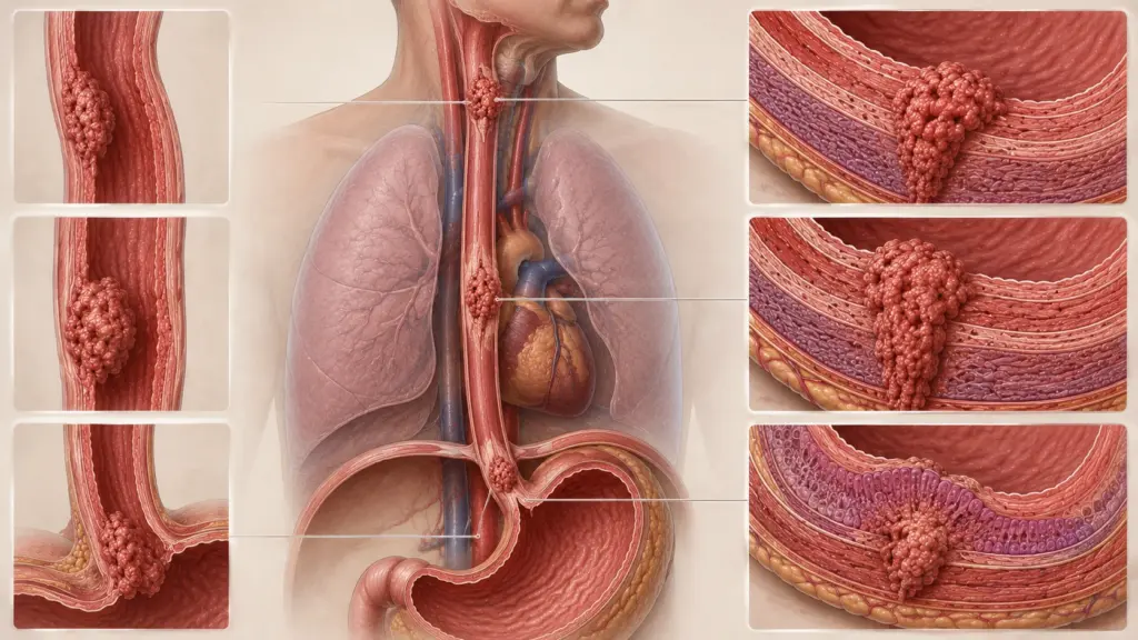
Esophageal cancer is a malignant tumor that originates in the mucosal layer of the esophagus. There are two main histological types: squamous cell carcinoma (SCC) and adenocarcinoma. SCC arises from squamous cells in the upper and middle sections of the esophagus; adenocarcinoma, on the other hand, develops from glandular cells in the lower esophagus and the gastroesophageal junction (GEJ) region. According to SEER data, the incidence of adenocarcinoma is increasing in the United States, while the incidence of SCC is decreasing.
According to IARC GLOBOCAN 2022 data, 511,054 new cases are diagnosed worldwide each year, and 445,391 people die from this disease. The global age-standardized incidence rate (ASIR) is 5.00 per 100,000, and the age-standardized mortality rate (ASMR) is 4.30 per 100,000. The highest rates are observed in East Africa (ASIR: 7.60), East Asia (ASIR: 7.60), and South Africa (ASIR: 6.30). The disease is three times more common in men than in women.
According to Assoc. Prof. Dr. Nilay Şengül, the most critical step in diagnosing esophageal cancer is to perform an endoscopic evaluation of patients presenting with dysphagia (difficulty swallowing). According to the protocol we follow at Memorial Şişli Hospital, an urgent upper gastrointestinal endoscopy is scheduled for every patient with difficulty swallowing lasting longer than 2–3 weeks. This approach increases the rate of early-stage diagnosis and broadens treatment options.
What Is Barrett’s Esophagus?
Barrett’s esophagus is a condition in which the normal squamous epithelium of the esophagus is replaced by intestinal-type columnar epithelium as a result of gastroesophageal reflux disease (GERD). According to UpToDate data, a diagnosis of Barrett’s esophagus requires the presence of salmon-colored mucosa at least 1 cm proximal to the gastroesophageal junction and confirmation of intestinal metaplasia via biopsy. Barrett’s esophagus is the only known precursor lesion of esophageal adenocarcinoma.
According to UpToDate data, the annual risk of cancer progression in nondysplastic Barrett’s esophagus ranges from 0.1% to 0.4%. The risk of cancer is at least 30 times higher than in the general population; however, the individual absolute risk is low. Male gender, advanced age, a long segment of Barrett’s mucosa, and a family history increase the risk of progression. According to data from Castle Biosciences, the annual risk of progression in Barrett’s with a long segment ≥3 cm is 0.22% (1/454), 0.03% (1/3,333) for short segments <3 cm, and 0.01% (1/10,000) for ultra-short segments <1 cm.
According to a Johns Hopkins study published in *Nature* in 2020, independent risk factors for progression in Barrett’s esophagus include age, abdominal obesity, heavy smoking (>30 pack-years), low-grade dysplasia (LGD), and caffeine consumption. Protective factors include statins, SSRIs, and calcium/vitamin D supplements.
Symptoms of Esophageal Cancer
Symptoms of esophageal cancer vary depending on the tumor’s location, size, and stage. In the early stages, symptoms may be mild and easily overlooked by patients.
- Difficulty swallowing (dysphagia)—the most common initial symptom; it begins with solid foods and progresses to liquids
- Pain or a burning sensation behind the chest
- Weight loss and loss of appetite
- Persistent cough and shortness of breath (in cases of tracheal or bronchial invasion)
- Hoarseness (compression of the recurrent laryngeal nerve)
- Regurgitation (backflow of food)
- Bloody vomiting or melena (black, tarry stools)
Esophageal Cancer Cough
In esophageal cancer, a cough may develop as a result of tumor invasion into the trachea or bronchi. Additionally, in patients with an esophagotracheal fistula (an abnormal connection between the esophagus and the airway), a chronic cough and recurrent lung infections may occur due to the leakage of contents from the esophagus into the airway. This condition is a serious complication in advanced-stage disease and requires urgent intervention.
What Causes Esophageal Cancer?
Different risk factors play a role in the development of esophageal cancer depending on the histological type. The causes of squamous cell carcinoma (SCC) and adenocarcinoma differ.
| Risk Factor | Associated Type | Relative Risk Increase |
|---|---|---|
| Smoking | SCC (strong), adenocarcinoma | SHK: ~5–10 times |
| Alcohol consumption | SHK (strong) | ~3–5 times |
| Barrett’s esophagus | Adenocarcinoma | ~30–40 times (low absolute risk) |
| Obesity (central) | Adenocarcinoma | ~2–3 times |
| Gastroesophageal reflux (GERD) | Adenocarcinoma | ~5–7 times |
| Achalasia | SHK | ~16–30 times |
| Plummer-Vinson syndrome | UES (upper esophagus) | High |
According to IARC GLOBOCAN 2022 data, the risk of esophageal cancer is approximately three times higher in men than in women. East Asia, East Africa, and South Africa are the regions with the highest incidence and mortality rates. China alone accounts for 43.8% of the global total. The risk of EEPC increases synergistically when tobacco and alcohol are used together.
Esophageal Cancer Mortality Rate
Esophageal cancer ranks sixth among cancer deaths worldwide. According to IARC GLOBOCAN 2022 data, the mortality-to-incidence ratio is high, with 445,391 deaths. This is largely due to the disease being diagnosed primarily at an advanced stage.
According to SEER data (2016–2022), the 5-year relative survival rate in the U.S. is 22.2%. Survival rates by stage are as follows:
| Stage / Status | Case Rate | 5-Year Relative Survival |
|---|---|---|
| Localized | 19 | 48.6% |
| Regional | 32 | 29.1% |
| Distant (metastatic) | 39 | 5.3% |
| Unknown stage | 10 | 15.9 |
According to SEER data, 39% of cases are diagnosed at the stage of distant metastasis; this is the primary reason for the low overall survival rate. According to NIH/NCBI data, the 5-year cause-specific survival rate for adenocarcinoma is 23.4%, with a median survival of 15 months; for squamous cell carcinoma (SCC), these rates are 18.9% and 10 months, respectively.
Stages of Esophageal Cancer
Esophageal cancer staging is performed according to the AJCC TNM system. Tumor depth (T), lymph node involvement (N), and distant metastasis (M) are assessed. According to SEER data, most cases are diagnosed at an advanced stage.
According to SEER educational modules, the 5-year survival rates by stage are as follows:
- Stage 0 (carcinoma in situ): Survival rate over 90%
- Stage I: 50–70% for distal tumors; 10–15% for proximal tumors
- Stage II: Over 25% in T1/T2 N+ cases; less than 25% in T3 cases
- Stage III: 15% for distal tumors
- Stage IV: Less than 5%
According to NIH/NCBI data, the hazard ratio (HR) for Stage IV tumors in adenocarcinoma is 3.58 (95% CI: 3.33–3.85) compared to Stage I; while in squamous cell carcinoma (SCC), it is 2.12 (95% CI: 1.94–2.32). The risk in patients who did not undergo surgery is 2.54 times higher than in those who did.
Esophageal Cancer Treatment
Esophageal cancer treatment is personalized based on the tumor stage, histological type, location, and the patient’s overall condition. According to NCCN and ESMO guidelines, a multidisciplinary approach is essential in treatment planning.
Surgical Treatment
Esophagectomy (removal of the esophagus) is the primary treatment for early-stage and localized advanced-stage disease. According to NIH/NCBI data, surgical techniques include transhiatal esophagectomy, transthoracic esophagectomy (Ivor Lewis procedure), and minimally invasive esophagectomy (MIE). Lymph node dissection is performed during surgery. A gastric conduit or colonic interposition is created to ensure gastrointestinal continuity after resection.
According to a 2024 study in JAMA Network Open, in stage II–III patients, neoadjuvant chemoradiotherapy (NCRT) plus surgery significantly improves survival compared to definitive chemoradiotherapy (DCRT) alone (AHR: 0.45; 95% CI: 0.44–0.46; p < 0.001). Perioperative chemotherapy (FLOT) also yielded similarly superior results (AHR: 0.33; 95% CI: 0.28–0.39; p < 0.001).
Chemoradiotherapy
CROSS study: Surgery following neoadjuvant carboplatin + paclitaxel + 41.4 Gy of radiation therapy significantly prolonged overall survival compared to surgery alone. This regimen has become the standard of care for esophageal and GEJ cancers.
FLOT4 Trial: Perioperative FLOT chemotherapy (docetaxel, oxaliplatin, leucovorin, 5-FU) improved overall survival in resectable gastric and GEJ adenocarcinomas compared to the ECF/ECX regimen. According to the 2024 NCCN guidelines, FLOT is a Category 1 recommendation.
CheckMate 577: In patients with residual pathological disease following neoadjuvant chemoradiotherapy, adjuvant nivolumab significantly prolonged progression-free survival.
Immunotherapy
Immune checkpoint inhibitors are among the standard treatment options for advanced esophageal cancer. Pembrolizumab and nivolumab can be used in combination with platinum-based chemotherapy or as monotherapy. The combination of nivolumab and ipilimumab has demonstrated efficacy in selected patients.
Esophageal Stenting
An esophageal stent is used for palliative purposes to treat dysphagia in patients with advanced or inoperable disease. The self-expandable metal stent (SEMS) is the most commonly used type. The stent opens the narrowing caused by the tumor, allowing the patient to continue eating by mouth. However, a stent is not a curative treatment; it is intended solely for symptom control. Complications include stent migration, obstruction, fistula formation, and pain.
Esophageal Cancer Surgery
Esophageal cancer surgery (esophagectomy) aims to completely remove the tumor and restore gastrointestinal continuity. The surgical technique is determined based on the tumor’s location and stage.
Surgical Techniques
Transthoracic esophagectomy (Ivor Lewis): Performed via an approach through the abdominal and thoracic cavities for tumors of the lower and middle esophagus. A gastric conduit is created, and an anastomosis is performed within the thoracic cavity. This technique is the most commonly used approach.
Transhiatal esophagectomy: Performed via approaches through the abdomen and neck for tumors of the upper esophagus. No thoracotomy is performed; therefore, the risk of pulmonary complications is lower. However, the field of view is limited.
Minimally invasive esophagectomy (MIE): This combines laparoscopic and thoracoscopic approaches. It offers the advantages of less bleeding, a shorter hospital stay, and faster recovery compared to open surgery. However, long-term oncological outcomes are comparable.
Risks of Esophageal Cancer Surgery
Esophagectomy is a major surgical procedure carrying a high risk of morbidity and mortality. According to NIH/NCBI data, the complication rate ranges from 40% to 60%. The main risks are:
- Anastomotic leak (5–20%): This is the most serious complication; it can lead to mediastinitis and sepsis
- Pulmonary complications (20–30%): Pneumonia, atelectasis, pleural effusion
- Cardiac arrhythmias (10–20%): Especially after thoracotomy
- Recurrent laryngeal nerve injury (5–10%): Causes hoarseness
- Chyleus and gastroparesis: Motility disorders of the gastric conduit
- Dumping syndrome: Early and late types; nausea, sweating, and palpitations after eating
Assoc. Prof. Dr. Şengül emphasizes that anastomotic leakage is the most serious and life-threatening complication following esophagectomy in clinical practice. For this reason, she notes that it is critical for the surgery to be performed at an experienced center by a high-volume surgeon.
Nutrition After Esophageal Cancer Surgery
Nutrition following esophagectomy directly impacts the patient’s long-term quality of life. Enteral or parenteral nutrition is administered during the first 1–2 weeks. Since the gastric conduit is narrowed, small, frequent meals are recommended. To prevent dumping syndrome, liquids and solid foods should be consumed separately. Vitamin and mineral (B12, iron, calcium, vitamin D) supplementation continues for life. Assoc. Prof. Dr. Şengül emphasizes that, in her clinical practice, regular follow-up with a nutritionist significantly improves patients’ quality of life after surgery.
| Phase | Nutritional Approach | Restrictions |
|---|---|---|
| First 1–2 Weeks | Enteral/parenteral nutrition | No oral feeding |
| Weeks 2–4 | Liquid and soft diet | Avoid solid foods |
| 1–3 months | Small, frequent meals | Separating liquids and solids |
| 3+ months | Transition to a normal diet | Precautions for Dumping Syndrome |
Clinical Assessment by Assoc. Prof. Dr. Nilay Şengül
In my clinical practice, I frequently observe the following: The vast majority (70–80%) of patients diagnosed with esophageal cancer are in an advanced stage at the time of diagnosis and are no longer candidates for surgery. In the cases we evaluated at Memorial Şişli Hospital, the primary reason for this is that early symptoms (mild dysphagia, heartburn) are dismissed as “reflux” or “aging.”
According to Assoc. Prof. Dr. Nilay Şengül, the most significant paradigm shift in esophageal cancer is the transition from a “surgery alone” approach to “neoadjuvant multimodal therapy + surgery.” The CROSS and FLOT4 studies have demonstrated that this approach significantly prolongs survival. In particular, the CheckMate 577 study has solidified the role of immunotherapy by demonstrating that adjuvant nivolumab prolongs progression-free survival in patients with residual disease following neoadjuvant therapy. Assoc. Prof. Dr. Şengül emphasizes that regular endoscopic surveillance in patients diagnosed with Barrett’s esophagus can increase the rate of early detection of adenocarcinoma.
From Diagnosis to Recovery: How Does the Process Proceed?
- Initial Examination and Evaluation: The patient’s symptoms (duration of dysphagia, weight loss, pain), lifestyle (smoking, alcohol, diet), and family history are assessed. During the physical examination, the lymph nodes, liver, and abdomen are examined. Assoc. Prof. Dr. Şengül emphasizes that in her clinical practice, dysphagia that begins with solid foods and progresses to liquids is the most important warning sign for esophageal cancer.
- Diagnostic Tests:
- Upper gastrointestinal endoscopy (EGD): The gold standard for diagnosis; it determines the tumor’s location, size, and extent; a biopsy is taken
- EUS (Endoscopic ultrasound): To assess tumor depth (T stage) and peritumoral lymph nodes
- CT (chest + abdomen + pelvis): For screening for distant metastases; liver, lung, adrenal, and bone metastases
- PET-CT: More sensitive than CT for detecting metastases; particularly useful for lymph node staging
- Laparoscopy: To assess peritoneal spread in tumors of the gastroesophageal junction (GEJ) and distal esophagus
- Staging and Treatment Planning: AJCC TNM staging is applied. An individualized treatment plan is developed by a multidisciplinary tumor board (medical oncology, radiation oncology, gastroenterology, thoracic surgery, radiology, and pathology). According to the 2024 NCCN guidelines, upfront surgery or neoadjuvant therapy is planned for stages I–IIA; neoadjuvant chemoradiotherapy or perioperative chemotherapy plus surgery is planned for stages IIB–III; and systemic therapy or palliative care is planned for stage IV.
- Treatment Implementation:
- Early-stage (T1–2 N0): Surgery or endoscopic resection (for mucosal tumors)
- Localized advanced stage (T3–4 N+): Neoadjuvant chemoradiotherapy (CROSS) or perioperative FLOT plus surgery
- Advanced/metastatic stage: Systemic chemotherapy plus immunotherapy; palliative radiation therapy or a stent (for dysphagia)
- Hospital Stay and Discharge: The hospital stay following esophagectomy is 7–14 days. With a minimally invasive approach, this duration may be reduced to 5–10 days. Enteral feeding (jejunostomy) may continue after discharge. Hospital stay is prolonged in patients who develop complications.
- Post-Discharge Recovery: A liquid or soft diet for the first 2–4 weeks; small, frequent meals for 1–3 months. Return to work is generally possible 6–12 weeks later. Measures to prevent dumping syndrome (separating liquids and solids, eating small meals) are continued for life. Vitamin and mineral supplements (B12, iron, calcium, D) are recommended for life. Follow-up appointments are scheduled every 3 months for the first 2 years, and every 6 months for the following 3 years.
Frequently Asked Questions
What helps with esophageal cancer?
Esophageal cancer treatment is personalized based on the stage and histological type. In early stages, surgery or endoscopic resection is performed; in localized advanced stages, neoadjuvant chemoradiotherapy (CROSS) or perioperative FLOT chemotherapy plus surgery is used; and in advanced stages, systemic chemotherapy plus immunotherapy is administered. Palliative treatment involves the use of esophageal stents, radiation therapy, and nutritional support. According to Assoc. Prof. Dr. Şengül, regular endoscopic surveillance in Barrett’s esophagus can facilitate the early diagnosis of adenocarcinoma.
At what age does esophageal cancer occur?
The incidence of esophageal cancer increases with age; the median age at diagnosis is between 60 and 70. However, this varies depending on the histological type: squamous cell carcinoma (SCC) can occur at younger ages (50–60), while adenocarcinoma is more common in patients aged 60–70. Adenocarcinoma associated with Barrett’s esophagus can develop at an earlier age in patients with a history of long-term reflux. It is three times more common in men than in women.
Who is at risk for esophageal cancer?
It is three times more common in men than in women. The risk of squamous cell carcinoma is high among smokers, heavy drinkers, and those with achalasia or Plummer-Vinson syndrome. The risk of adenocarcinoma, on the other hand, increases in individuals with Barrett’s esophagus, central obesity, gastroesophageal reflux disease (GERD), and smokers. The incidence of esophageal squamous cell carcinoma (ESC) is higher among individuals of East Asian, East African, and South African descent; meanwhile, the incidence of adenocarcinoma is rising in Western countries due to the increasing prevalence of obesity.
Does esophageal cancer recur?
Yes, it can recur. Local recurrence may develop at the resection site or along the anastomosis line. Distant recurrence is commonly seen in the liver, lungs, bones, and adrenal glands. The risk of recurrence varies depending on the stage, lymph node involvement, and surgical margins. Adjuvant therapy (chemotherapy, radiation therapy, immunotherapy) can reduce the risk of recurrence. In cases of recurrence or refractory disease, systemic treatment or participation in clinical trials is considered.
How is esophageal cancer diagnosed?
A definitive diagnosis is made by taking a biopsy during endoscopy (EGD). Endoscopic ultrasound (EUS) evaluates tumor depth and lymph node involvement. CT is used to screen for distant metastases. PET-CT is more sensitive than CT for detecting metastases. Laparoscopy is helpful in assessing peritoneal spread in GEJ tumors. Endoscopic ultrasound-guided fine-needle aspiration biopsy (EUS-FNA) can be used for lymph node staging.
Is there a treatment for esophageal cancer?
Yes, there is a treatment. In early stages (Stages 0–I), a cure can be achieved with surgery or endoscopic resection. In localized advanced stages (Stages II–III), 5-year survival rates can reach 40–50% with neoadjuvant chemoradiotherapy (CROSS) or perioperative FLOT chemotherapy plus surgery. In advanced stages (Stage IV), the disease can be controlled with systemic chemotherapy and immunotherapy; however, a cure is not possible. Palliative treatments (stents, radiation therapy) help manage symptoms.
How is esophageal cancer surgery performed?
Esophagectomy is the surgical removal of the esophagus. Transthoracic (Ivor Lewis), transhiatal, or minimally invasive (MIE) approaches are used. During surgery, the tumor and surrounding lymph nodes are removed; gastrointestinal continuity is restored using a gastric conduit or colonic interposition. The surgery takes 4–8 hours, and the hospital stay is 7–14 days. It is critical that the procedure be performed at an experienced center by a high-volume surgeon.
How long does esophageal cancer surgery take?
The duration of an esophagectomy varies between 4 and 8 hours, depending on the technique used and the complexity of the tumor. The transthoracic (Ivor Lewis) procedure typically takes 5–7 hours. Minimally invasive esophagectomy (MIE) may take longer than open surgery; however, it offers the advantages of less bleeding and a faster recovery. In cases where complications arise, the surgery may take longer.
Is esophageal cancer dangerous?
Yes, it is a cancer with a high mortality rate. According to IARC GLOBOCAN 2022 data, it ranks sixth among cancer deaths worldwide. According to SEER data, the 5-year relative survival rate is 22.2%. Thirty-nine percent of cases are diagnosed at the stage of distant metastasis; in such cases, the 5-year survival rate drops to 5.3%. Early diagnosis and multimodal treatment significantly improve the prognosis. The CROSS study has demonstrated that the combination of neoadjuvant therapy and surgery prolongs survival.
Can esophageal cancer be detected through blood tests?
Blood tests do not directly establish a diagnosis; however, they can be helpful. Anemia (resulting from bloody vomiting or melena), abnormal liver function tests (indicating liver metastases), elevated LDH, and low albumin levels may suggest the presence of metastatic disease. Tumor markers (CEA, CA 19-9, SCC antigen) are used for monitoring, not for diagnosis. A definitive diagnosis is made through endoscopy and biopsy.
Is esophageal cancer fatal?
In advanced stages, yes, it can be fatal. According to SEER data, the 5-year relative survival rate is 22.2%. It is 48.6% for localized disease, 29.1% for regionally advanced disease, and 5.3% for disease with distant metastases. According to IARC GLOBOCAN 2022 data, 445,391 people worldwide die from this disease. However, in early stages (Stages 0–I), the 5-year survival rate can reach 50–90% with surgery. Therefore, early diagnosis is of critical importance.
What is the life expectancy for esophageal cancer?
Life expectancy varies depending on the stage and response to treatment. The 5-year survival rate is 48.6% for localized disease, 29.1% for regionally advanced disease, and 5.3% for disease with distant metastases. According to NIH/NCBI data, the median survival is 15 months for adenocarcinoma and 10 months for squamous cell carcinoma (SCC). In the CROSS study, median survival was significantly prolonged with neoadjuvant chemoradiotherapy plus surgery compared to surgery alone. Perioperative FLOT chemotherapy plus surgery has similarly yielded superior results.
Which department treats esophageal cancer?
A multidisciplinary approach is used in the diagnosis and treatment of esophageal cancer. The physician who makes the diagnosis and performs the initial evaluation is typically a gastroenterologist. Teams from medical oncology, radiation oncology, thoracic surgery, and gastroenterology are involved in treatment planning. According to NCCN and ESMO guidelines, all esophageal cancer patients should be evaluated at an experienced center. Endoscopic treatments (EMR, ESD) are planned by a gastroenterologist; surgery is performed by a thoracic surgeon; and systemic therapy is planned by a medical oncologist.
Esophageal cancer is an aggressive malignancy that represents a significant cancer burden worldwide and is often diagnosed at an advanced stage. According to IARC GLOBOCAN 2022 data, it ranks sixth among cancer deaths, with 511,054 new cases and 445,391 deaths. Survival rates can be significantly improved with early diagnosis and treatment. The CROSS and FLOT4 studies have demonstrated that the combination of neoadjuvant multimodal therapy and surgery prolongs survival. Regular endoscopic surveillance in patients diagnosed with Barrett’s esophagus can increase the rate of early detection of adenocarcinoma.
To discuss your personal situation regarding esophageal cancer and have your questions answered, you can consult with Assoc. Prof. Dr. Nilay Şengül. To schedule an appointment at Memorial Şişli Hospital, please call 444 7 888.
Sources
- IARC GLOBOCAN 2022, Esophageal Cancer Fact Sheet — https://gco.iarc.who.int/media/globocan/factsheets/cancers/6-oesophagus-fact-sheet.pdf
- PMC/NIH, "Global esophageal cancer epidemiology in 2022 and predictions for 2050" — https://pmc.ncbi.nlm.nih.gov/articles/PMC11706580/
- SEER Cancer Stat Facts: Esophageal Cancer — https://seer.cancer.gov/statfacts/html/esoph.html
- NIH/NCBI StatPearls, "Esophageal Cancer" — https://www.ncbi.nlm.nih.gov/books/NBK459267/
- UpToDate, "Barrett's esophagus: Surveillance and management" — https://www.uptodate.com/contents/barretts-esophagus-surveillance-and-management/print
- Nature/Scientific Reports, "Risk Factors for Progression of Barrett's Esophagus to High-Grade Dysplasia and Esophageal Adenocarcinoma" — https://www.nature.com/articles/s41598-020-61874-7
- JAMA Network Open, "Survival and Treatment Patterns in Stage II to III Esophageal Cancer" — https://jamanetwork.com/journals/jamanetworkopen/fullarticle/2825137
- PMC/NIH, "Esophageal Cancer: An Updated Surveillance, Epidemiology, and End Results Database Analysis" — https://pmc.ncbi.nlm.nih.gov/articles/PMC7141161/
- Castle Biosciences, "Barrett's Esophagus Cancer Risk" — https://castlebiosciences.com/patient-information/gastroenterology/barretts-esophagus/cancer-risk
- SEER Training Modules, "Five-Year Survival Rates" — https://training.seer.cancer.gov/ugi/intro/survival.html